Lipotoxicity of the pancreatic beta-cell is associated with glucose-dependent esterification of fatty acids into neutral lipids - PubMed (original) (raw)
Lipotoxicity of the pancreatic beta-cell is associated with glucose-dependent esterification of fatty acids into neutral lipids
I Briaud et al. Diabetes. 2001 Feb.
Abstract
Prolonged exposure of isolated islets to supraphysiologic concentrations of palmitate decreases insulin gene expression in the presence of elevated glucose levels. This study was designed to determine whether or not this phenomenon is associated with a glucose-dependent increase in esterification of fatty acids into neutral lipids. Gene expression of sn-glycerol-3-phosphate acyltransferase (GPAT), diacylglycerol acyltransferase (DGAT), and hormone-sensitive lipase (HSL), three key enzymes of lipid metabolism, was detected in isolated rat islets. Their levels of expression were not affected after a 72-h exposure to elevated glucose and palmitate. To determine the effects of glucose on palmitate-induced neutral lipid synthesis, isolated rat islets were cultured for 72 h with trace amounts of [14C]palmitate with or without 0.5 mmol/l unlabeled palmitate, at 2.8 or 16.7 mmol/l glucose. Glucose increased incorporation of [14C]palmitate into complex lipids. Addition of exogenous palmitate directed lipid metabolism toward neutral lipid synthesis. As a result, neutral lipid mass was increased upon prolonged incubation with elevated palmitate only in the presence of high glucose. The ability of palmitate to increase neutral lipid synthesis in the presence of high glucose was concentration-dependent in HIT cells and was inversely correlated to insulin mRNA levels. 2-Bromopalmitate, an inhibitor of fatty acid mitochondrial beta-oxidation, reproduced the inhibitory effect of palmitate on insulin mRNA levels. In contrast, palmitate methyl ester, which is not metabolized, and the medium-chain fatty acid octanoate, which is readily oxidized, did not affect insulin gene expression, suggesting that fatty-acid inhibition of insulin gene expression requires activation of the esterification pathway. These results demonstrate that inhibition of insulin gene expression upon prolonged exposure of islets to palmitate is associated with a glucose-dependent increase in esterification of fatty acids into neutral lipids.
Figures
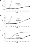
FIG. 1
Detection of GPAT (A), DGAT (B), and HSL (C) mRNAs by RT-PCR using Taqman technology in isolated rat islets. ΔRn designates changes in fluorescence emission. Representative amplification plots were from experiments performed in triplicate on two separate occasions.
FIG. 2
Effects of prolonged exposure to glucose (Gluc.) and palmitate (Palm.) on insulin (A), GPAT (B), DGAT (C), and HSL (D) mRNA levels in isolated rat islets. Isolated islets were incubated in the absence or presence of 0.5 mmol/l palmitate and 2.8 or 16.7 mmol/l glucose for 72 h. Insulin (n = 8), GPAT (n = 3), DGAT (n = 7), and HSL (n = 3) mRNA levels were determined by fluorescence-based RT-PCR as described in
research design and methods
. Results (means ± SE) are expressed as the ratio between the signal corresponding to the gene of interest and the signal for β-actin mRNA. *P < 0.01.
FIG. 3
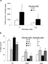
Effects of palmitate and glucose on incorporation of [14C] exogenous palmitate in isolated rat islets. Isolated rat islets were incubated with 5 µmol/l [U-14C]palmitate in the absence or presence of 0.5 mmol/l unlabeled palmitate and 2.8 or 16.7 mmol/l glucose for 72 h. Extracted lipids were analyzed by TLC. For each lane, the lipid bands were scraped and counted. Results are means ± SE of seven to nine separate experiments. A: Effects of glucose on the total number of counts incorporated from labeled palmitate into complex lipids (PL + DAG + DAG). The effect of palmitate cannot be ascertained because of the dilution of the specific activity of the tracer in the presence of unlabeled palmitate. Indeed, the absolute number of counts is artificially lower in the presence of 0.5 mmol/l unlabeled palmitate. *P < 0.05. B: Effects of glucose and palmitate on the distribution of the counts into each complex lipid fraction. Results are expressed as percent of total counts recovered from each lane. *P < 0.05; **P < 0.01.
FIG. 4
Effects of palmitate and glucose on TAG content in isolated rat islets. Isolated rat islets were incubated in the absence or presence of 0.5 mmol/l palmitate and 2.8 or 16.7 mmol/l glucose for 72 h. TAG content was measured as described in
research design and methods
. Results are means ± SE of three to six separate experiments. *P < 0.05; **P < 0.01.
FIG. 5
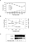
Dose-dependent effects of palmitate on lipid partitioning, TAG content, and insulin mRNA levels in HIT-T15 cells. HIT-T15 cells were incubated for 72 h in RPMI 1640 containing 11.1 mmol/l glucose with increasing concentrations of palmitate. A: The culture medium contained 5 µmol/l [U-14C]palmitate. Lipids were extracted and analyzed by TLC. For each lane, the lipid bands were scraped and counted. Results are means ± SE of three separate experiments and are expressed as percent of total counts recovered from each lane. All points at and above 0.25 mmol/l are significantly different from the control (0 palmitate) for PL, TAG, and DAG. CE, cholesterol ester. B: Inverse correlation between TAG content and insulin mRNA levels. Results are expressed as means ± SE of five or six separate experiments. C: Representative Northern blot of insulin mRNA.
FIG. 6
Effects of fatty acids on insulin mRNA level in HIT-T15 cells. HIT-T15 cells were cultured for 24 h in the presence of 0.5 mmol/l palmitate, 2.5 mmol/l octanoate, 0.5 mmol/l palmitate methyl ester, or 0.5 mmol/l 2-bromopalmitate. The control condition contained an equivalent amount of BSA. Insulin mRNA was measured by Northern analysis and was normalized to β-actin mRNA. Results are means ± SE of three separate experiments and are expressed as percent control. A representative Northern blot is shown on the right.
Similar articles
- Inhibition of insulin gene expression by long-term exposure of pancreatic beta cells to palmitate is dependent on the presence of a stimulatory glucose concentration.
Jacqueminet S, Briaud I, Rouault C, Reach G, Poitout V. Jacqueminet S, et al. Metabolism. 2000 Apr;49(4):532-6. doi: 10.1016/s0026-0495(00)80021-9. Metabolism. 2000. PMID: 10778881 - Regulatory Role of Fatty Acid Metabolism on Glucose-Induced Changes in Insulin and Glucagon Secretion by Pancreatic Islet Cells.
Tamarit-Rodriguez J. Tamarit-Rodriguez J. Int J Mol Sci. 2024 May 31;25(11):6052. doi: 10.3390/ijms25116052. Int J Mol Sci. 2024. PMID: 38892240 Free PMC article. Review. - Genetic manipulations of fatty acid metabolism in beta-cells are associated with dysregulated insulin secretion.
Eto K, Yamashita T, Matsui J, Terauchi Y, Noda M, Kadowaki T. Eto K, et al. Diabetes. 2002 Dec;51 Suppl 3:S414-20. doi: 10.2337/diabetes.51.2007.s414. Diabetes. 2002. PMID: 12475784 Review.
Cited by
- Identification and Characterization of Circular Intronic RNAs Derived from Insulin Gene.
Das D, Das A, Sahu M, Mishra SS, Khan S, Bejugam PR, Rout PK, Das A, Bano S, Mishra GP, Raghav SK, Dixit A, Panda AC. Das D, et al. Int J Mol Sci. 2020 Jun 17;21(12):4302. doi: 10.3390/ijms21124302. Int J Mol Sci. 2020. PMID: 32560282 Free PMC article. - Lipotoxic Impairment of Mitochondrial Function in β-Cells: A Review.
Römer A, Linn T, Petry SF. Römer A, et al. Antioxidants (Basel). 2021 Feb 15;10(2):293. doi: 10.3390/antiox10020293. Antioxidants (Basel). 2021. PMID: 33672062 Free PMC article. Review. - Adverse physicochemical properties of tripalmitin in beta cells lead to morphological changes and lipotoxicity in vitro.
Moffitt JH, Fielding BA, Evershed R, Berstan R, Currie JM, Clark A. Moffitt JH, et al. Diabetologia. 2005 Sep;48(9):1819-29. doi: 10.1007/s00125-005-1861-9. Epub 2005 Aug 11. Diabetologia. 2005. PMID: 16094531 - Regulation of the insulin gene by glucose and fatty acids.
Poitout V, Hagman D, Stein R, Artner I, Robertson RP, Harmon JS. Poitout V, et al. J Nutr. 2006 Apr;136(4):873-6. doi: 10.1093/jn/136.4.873. J Nutr. 2006. PMID: 16549443 Free PMC article. Review. - Mechanisms involved in the cytotoxic and cytoprotective actions of saturated versus monounsaturated long-chain fatty acids in pancreatic beta-cells.
Diakogiannaki E, Dhayal S, Childs CE, Calder PC, Welters HJ, Morgan NG. Diakogiannaki E, et al. J Endocrinol. 2007 Aug;194(2):283-91. doi: 10.1677/JOE-07-0082. J Endocrinol. 2007. PMID: 17641278 Free PMC article.
References
- Unger RH. Lipotoxicity in the pathogenesis of obesity-dependent NIDDM: genetic and clinical implications. Diabetes. 1995;44:863–870. - PubMed
- McGarry JD, Dobbins RL. Fatty acids, lipotoxicity and insulin secretion. Diabetologia. 1999;42:128–138. - PubMed
- Jacqueminet S, Briaud I, Rouault C, Reach G, Poitout V. Inhibition of insulin gene expression by long-term exposure of pancreatic beta-cells to palmitate is dependent upon the presence of a stimulatory glucose concentration. Metabolism. 2000;49:532–536. - PubMed
- Gremlich S, Bonny C, Waeber G, Thorens B. Fatty acids decrease IDX-1 expression in rat pancreatic islets and reduce GLUT2, glucokinase, insulin, and somatostatin levels. J Biol Chem. 1997;272:30261–30269. - PubMed
- Ritz-Laser B, Meda P, Constant I, Klages N, Charollais A, Morales A, Magnan C, Ktorza A, Philippe J. Glucose-induced preproinsulin gene expression is inhibited by the free-fatty acid palmitate. Endocrinology. 1999;140:4005–4014. - PubMed
Publication types
MeSH terms
Substances
LinkOut - more resources
Full Text Sources
Research Materials


