FGF-2 regulation of neurogenesis in adult hippocampus after brain injury - PubMed (original) (raw)
FGF-2 regulation of neurogenesis in adult hippocampus after brain injury
S Yoshimura et al. Proc Natl Acad Sci U S A. 2001.
Abstract
Fibroblast growth factor-2 (FGF-2) promotes proliferation of neuroprogenitor cells in culture and is up-regulated within brain after injury. Using mice genetically deficient in FGF-2 (FGF-2(-/-) mice), we addressed the importance of endogenously generated FGF-2 on neurogenesis within the hippocampus, a structure involved in spatial, declarative, and contextual memory, after seizures or ischemic injury. BrdUrd incorporation was used to mark dividing neuroprogenitor cells and NeuN expression to monitor their differentiation into neurons. In the wild-type strain, hippocampal FGF-2 increased after either kainic acid injection or middle cerebral artery occlusion, and the numbers of BrdUrd/NeuN-positive cells significantly increased on days 9 and 16 as compared with the controls. In FGF-2(-/-) mice, BrdUrd labeling was attenuated after kainic acid or middle cerebral artery occlusion, as was the number of neural cells colabeled with both BrdUrd and NeuN. After FGF-2(-/-) mice were injected intraventricularly with a herpes simplex virus-1 amplicon vector carrying FGF-2 gene, the number of BrdUrd-labeled cells increased significantly to values equivalent to wild-type littermates after kainate seizures. These results indicate that endogenously synthesized FGF-2 is necessary and sufficient to stimulate proliferation and differentiation of neuroprogenitor cells in the adult hippocampus after brain insult.
Figures
Figure 1
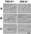
BrdUrd-positive cells in the medial dentate gyrus of FGF-2+/+ and FGF-2−/− mice after brain injury. After kainic acid injection, MCAO or no injury (control), BrdUrd was injected 6, 7, and 8 days later (to label dividing cells), and animals were killed on day 9. Few BrdUrd-labeled cells were detected in the untreated FGF-2+/+ and FGF-2−/− mice (A). After kainic acid injection (B) or MCAO (C), greater cell proliferation was detected in a region corresponding to the subgranular layer in FGF-2+/+ littermates. Immunohistochemistry was performed on free-floating 50-μm coronal sections pretreated by denaturing DNA (see Materials and Methods). (Scale bar, 100 μm.)
Figure 2
Quantification of BrdUrd-positive cells in dentate gyrus after kainic acid injection (A) or MCAO (B) in FGF-2+/+ (empty bars) and FGF-2−/− mice (filled bars). The number of BrdUrd-labeled cells was counted on days 9 or 16 after injury (see Materials and Methods). Note the large early increase in labeled cells after kainic acid and MCAO, which was attenuated at both time points in the FGF-2−/− mice. The ordinate scale reflects the counts in the total dentate gyrus (means + SD, n = 6 per group). +,P < 0.05 compared with sham control. *,P < 0.01 compared with FGF-2+/+ littermates on the same day of death.
Figure 3
Neuronal and glial identity of newly divided cells in the dentate gyrus before (A) and after (B and_C_) kainic acid treatment in FGF-2+/+ and FGF-2−/− mice. Brain sections (50 μm) were stained for BrdUrd immunoreactivity (FITC, green) and cell-specific markers (cy3, red) for either neurons (NeuN) (A and B) or glia (glial fibrillary acidic protein) (C) and examined by confocal microscopy. BrdUrd was injected on days 6, 7, and 8 after injury or no injury (control) and animals were killed on day 35 after kainic acid injection. Cells with colocalization of BrdUrd and NeuN (yellow nuclei) are more numerous after kainic acid treatment in FGF-2+/+ as compared with FGF-2−/− mice, and nearly all BrdUrd-positive cells were also NeuN positive. Colocalization of glial fibrillary acidic protein and BrdUrd labeling was rarely observed in either FGF-2+/+ or control brains. (Scale bar = 50 μm.)
Figure 4
FGF-2 gene transfer via HSV-1 amplicon vector increases neurogenesis in the dentate gyrus of FGF-2+/+ and FGF-2−/− mice with and without insult. HSV-1/empty and HSV-1/mFGF-2 were injected stereotactically into the lateral cerebroventricle, and kainic acid was injected or not injected the next day. BrdUrd then was injected as described (see Materials and Methods), and animals were killed on day 9 after kainic acid treatment. Proliferative activity was determined by counting BrdUrd-labeled cells, and data are expressed as total number of positive cells per dentate gyrus. Injection of HSV-1/empty virus amplicon vector did not modify the response observed previously to kainic acid; however, the FGF-2-bearing vector dramatically increase BrdUrd labeling in the FGF-2−/− mice to levels nearly equivalent to FGF-2+/+ mice (means + SD,n = 4 per group). *, P < 0.01 compared with the group injected with HSV-1/empty virus amplicon vector.
Similar articles
- FGF-2 regulates neurogenesis and degeneration in the dentate gyrus after traumatic brain injury in mice.
Yoshimura S, Teramoto T, Whalen MJ, Irizarry MC, Takagi Y, Qiu J, Harada J, Waeber C, Breakefield XO, Moskowitz MA. Yoshimura S, et al. J Clin Invest. 2003 Oct;112(8):1202-10. doi: 10.1172/JCI16618. J Clin Invest. 2003. PMID: 14561705 Free PMC article. - [Therapeutic neurogenesis for CNS disorders].
Yoshimura S, Sakai N. Yoshimura S, et al. Nihon Ronen Igakkai Zasshi. 2004 Jan;41(1):58-60. doi: 10.3143/geriatrics.41.58. Nihon Ronen Igakkai Zasshi. 2004. PMID: 14999916 Japanese. - Human FGF-1 gene delivery protects against quinolinate-induced striatal and hippocampal injury in neonatal rats.
Hossain MA, Fielding KE, Trescher WH, Ho T, Wilson MA, Laterra J. Hossain MA, et al. Eur J Neurosci. 1998 Aug;10(8):2490-9. Eur J Neurosci. 1998. PMID: 9767380 - Enlarged infarct volume and loss of BDNF mRNA induction following brain ischemia in mice lacking FGF-2.
Kiprianova I, Schindowski K, von Bohlen und Halbach O, Krause S, Dono R, Schwaninger M, Unsicker K. Kiprianova I, et al. Exp Neurol. 2004 Oct;189(2):252-60. doi: 10.1016/j.expneurol.2004.06.004. Exp Neurol. 2004. PMID: 15380477 - Expression and functions of fibroblast growth factor 2 (FGF-2) in hippocampal formation.
Zechel S, Werner S, Unsicker K, von Bohlen und Halbach O. Zechel S, et al. Neuroscientist. 2010 Aug;16(4):357-73. doi: 10.1177/1073858410371513. Epub 2010 Jun 25. Neuroscientist. 2010. PMID: 20581332 Review.
Cited by
- Hydrogel in the Treatment of Traumatic Brain Injury.
Li S, Xu J, Qian Y, Zhang R. Li S, et al. Biomater Res. 2024 Sep 26;28:0085. doi: 10.34133/bmr.0085. eCollection 2024. Biomater Res. 2024. PMID: 39328790 Free PMC article. Review. - FGF2 Functions in H2S's Attenuating Effect on Brain Injury Induced by Deep Hypothermic Circulatory Arrest in Rats.
Zhu YX, Yang Q, Zhang YP, Liu ZG. Zhu YX, et al. Mol Biotechnol. 2024 Dec;66(12):3526-3537. doi: 10.1007/s12033-023-00952-3. Epub 2023 Nov 2. Mol Biotechnol. 2024. PMID: 37919618 Free PMC article. - Fibroblast growth factor 2.
Nickle A, Ko S, Merrill AE. Nickle A, et al. Differentiation. 2024 Sep-Oct;139:100733. doi: 10.1016/j.diff.2023.10.001. Epub 2023 Oct 12. Differentiation. 2024. PMID: 37858405 Review. - Transcriptome and methylome of the supraoptic nucleus provides insights into the age-dependent loss of neuronal plasticity.
Thompson D, Odufuwa AE, Brissette CA, Watt JA. Thompson D, et al. Front Aging Neurosci. 2023 Aug 30;15:1223273. doi: 10.3389/fnagi.2023.1223273. eCollection 2023. Front Aging Neurosci. 2023. PMID: 37711995 Free PMC article. - Enriched environment-induced neuroplasticity in ischemic stroke and its underlying mechanisms.
Han PP, Han Y, Shen XY, Gao ZK, Bi X. Han PP, et al. Front Cell Neurosci. 2023 Jul 7;17:1210361. doi: 10.3389/fncel.2023.1210361. eCollection 2023. Front Cell Neurosci. 2023. PMID: 37484824 Free PMC article. Review.
References
- Altman J, Das G D. J Comp Neurol. 1965;124:319–335. - PubMed
- Kempermann G, Kuhn H G, Gage F H. Nature (London) 1997;386:493–495. - PubMed
Publication types
MeSH terms
Substances
Grants and funding
- 5 P50 NS10828/NS/NINDS NIH HHS/United States
- MH60587/MH/NIMH NIH HHS/United States
- P50 NS010828/NS/NINDS NIH HHS/United States
- P01 NS024279/NS/NINDS NIH HHS/United States
- NS24279/NS/NINDS NIH HHS/United States
LinkOut - more resources
Full Text Sources
Other Literature Sources
Molecular Biology Databases


