Cyclin-dependent kinase inhibitor p27(Kip1) is required for mouse mammary gland morphogenesis and function - PubMed (original) (raw)
Cyclin-dependent kinase inhibitor p27(Kip1) is required for mouse mammary gland morphogenesis and function
R S Muraoka et al. J Cell Biol. 2001.
Abstract
We have studied the role of the cyclin-dependent kinase (Cdk) inhibitor p27(Kip1) in postnatal mammary gland morphogenesis. Based on its ability to negatively regulate cyclin/Cdk function, loss of p27 may result in unrestrained cellular proliferation. However, recent evidence about the stabilizing effect of p27 on cyclin D1-Cdk4 complexes suggests that p27 deficiency might recapitulate the hypoplastic mammary phenotype of cyclin D1-deficient animals. These hypotheses were investigated in postnatal p27-deficient (p27(-/-)), hemizygous (p27(+/)-), or wild-type (p27(+/+)) mammary glands. Mammary glands from p27(+/)- mice displayed increased ductal branching and proliferation with delayed postlactational involution. In contrast, p27(-/-) mammary glands or wild-type mammary fat pads reconstituted with p27(-/-) epithelium produced the opposite phenotype: hypoplasia, low proliferation, decreased ductal branching, impaired lobuloalveolar differentiation, and inability to lactate. The association of cyclin D1 with Cdk4, the kinase activity of Cdk4 against pRb in vitro, the nuclear localization of cyclin D1, and the stability of cyclin D1 were all severely impaired in p27(-/-) mammary epithelial cells compared with p27(+/+) and p27(+/-) mammary epithelial cells. Therefore, p27 is required for mammary gland development in a dose-dependent fashion and positively regulates cyclin D-Cdk4 function in the mammary gland.
Figures
Figure 1
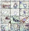
p27 is expressed in the mammary epithelium at all stages of postnatal morphogenesis. (A) Western blot analysis detecting p27 protein in p27 +/+ mammary glands harvested at the indicated stages of mammary gland development: pregnancy at 16.5 dpc; lactation at postnatal day 10; involution at 3 dpfw. Molecular weights (in kD) are indicated at left. (B–G) Immunohistochemical analysis of p27 expression in no. 4 intact virgin mammary glands harvested from 6-wk-old females or wild-type mammary fat pads reconstituted with tissue of the indicated genotype at 5 wk after transplantation. Arrowhead indicates nuclear localization of p27. Arrow in G indicates host vessel stained positive for p27. Panels shown are representative of results obtained for n = 3 per genotype, including reconstitutions.
Figure 4
Proliferation is increased in p27 +/− but decreased in p27− /− mammary epithelium. (A and B) Immunohistochemical detection of BrdU incorporation in mammary glands of 5-wk-old virgin mice (n = 6 per genotype). TEBs and ducts are shown. Genotypes are indicated. (C) BrdU incorporation into nuclei of wild-type mammary glands transplanted with p27 +/+, p27 +/−, or p27− /− mammary tissue. (D) BrdU incorporation into purified PMECs harvested from virgin mice of the indicated genotypes (n = 3 per genotype). (E) Quantification of BrdU incorporation in TEBs and in PMECs. The average number of BrdU-positive epithelial cells per single randomly chosen 400× field was determined. The proliferative index was calculated as follows: ([number of BrdU-labeled cells]/[total number of cells]) × 100. Each value represents the average of 10 fields from each of 6 individually analyzed mammary glands per genotype. (F) Flow cytometric analysis of propidium iodide–stained PMEC nuclei from virgin p27 +/+, p27 +/−, or p27− /− mice. Results are shown as the mean SEM of six mice per genotype. Bars, 25 μm.
Figure 2
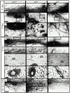
Altered ductal growth and branching in p27 +/− and p27− /− mammary glands. (A–E) Whole mounts of virgin no. 4 mammary glands at 21, 35, and 70 d. Asterisk denotes lymph nodes. All samples were photographed at the same magnification. Boxed areas (B and D) are shown enlarged directly below each panel (C and E) to show detail. Arrows indicate TEBs. (F–G) Hematoxylin and eosin–stained sections of mammary glands from virgin mice of each genotype at 70 d are shown. (H) Whole mount staining of virgin mammary glands from 240-d-old mice treated for 60 d with slow release estrogen–progesterone pellets. Arrowhead denotes area of focal hyperplasia. Panels shown are representative of results obtained for n = 3 per genotype. (I) Quantification of the number of TEBs per mammary gland at 35 d of age (n = 10 per genotype, see Fig. 2 B) and the number of epithelial structures per 100× field of hematoxylin and eosin–stained section of mammary gland from mice at 70 d of age (n = 10 fields per each of 6 glands per genotype, see Fig. 2 F). Numbers are presented as the average plus or minus the SEM; *P < 0.003, **P < 0.0001, # P < 0.03, @ P < 0.01, Student's t test. Bars: (A, B, D, and H) 400 μm; (C, E, and F) 100 μm; (G) 25 μm.
Figure 3
The morphological effects of p27 haploinsufficiency and of p27 deficiency are epithelial autonomous. (A and B) Whole mounts of wild-type mammary glands reconstituted with p27 +/+, p27+/−, or p27− /− tissue 5 wk after transplantation. Boxed areas are enlarged to show detail. Arrows indicate TEBs. (C and D) Primary mammary epithelial cells from virgin p27 +/+, p27 +/−, or p27− /− females (n = 3 mice per genotype) cultured in Matrigel. Representative photomicrographs shown were captured at 400× magnification (C) or at 100× (D) after 5 d; n > 20 colonies/genotype. Bars: (A) 400 μm; (B) 100 μm.
Figure 5
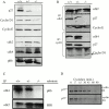
Loss of cyclin D1–Cdk4 activity and decreased cyclin D1 stability in p27 −/− mammary glands. (A) Western blot analysis of whole mammary gland extracts from virgin mice of each genotype was used to examine the content of p27, cyclin D1, cyclin E, Cdk2, Cdk4, and Rb. (B) IP using Cdk2 or cyclin D1 antibodies was performed on mammary gland extracts from mice of each genotype. Immunoprecipitates were subjected to Western blot using antibodies against p27, cyclin E, cyclin D1, Cdk2, or Cdk4 as indicated. (C) Kinase activity of Cdk4 and Cdk2 precipitates pRb (46 kD) or HH1 as indicated in Materials and Methods. (D) p27 +/+ or p27 −/− PMECs were cultured for 0–90 min in the presence of cycloheximide (5 μg/ml). Whole cell extracts were analyzed for the presence of cyclin D1 by Western blot analysis.
Figure 6
Impaired nuclear localization of cyclin D1 in the absence of p27. (A) Immunohistochemical detection of cyclin D1 protein in 6-wk-old virgin p27 +/+, p27 +/−, or p27− /− glands. (B) Quantification of the total number of cyclin D1–positive nuclei.
Figure 6
Impaired nuclear localization of cyclin D1 in the absence of p27. (A) Immunohistochemical detection of cyclin D1 protein in 6-wk-old virgin p27 +/+, p27 +/−, or p27− /− glands. (B) Quantification of the total number of cyclin D1–positive nuclei.
Figure 8
Mammary glands reconstituted with p27− /− epithelium display impaired function during lactation. (A–D) Hematoxylin and eosin staining of glands taken postpartum at 10 d (for intact glands) or 2 h (for reconstituted glands). Upper row shows low magnification (A–D); bottom row shows high magnification (E–H). (I and J) Whole mammary glands from p27 +/+ and p27− /− mice were cultured for 10 d in the presence of mammogenic hormones (described in Materials and Methods) to determine the ability of p27− /− glands to differentiate within the context of p27− /− stroma. Whole mounts are shown at 1 and 10 d of culture. Representative panels are shown; n = 10 per genotype. (K) Western blot analyses were used to examine the content of β-casein (β-cas), α-lactalbumin (α-LA), keratin-14 (K14), Stat5a, and phospho-Stat5 proteins in virgin p27 +/+ and lactating p27 +/+ glands or lactating wild-type glands reconstituted with p27− /− or p27 +/+ epithelium at 2 h postpartum. Results are representative of three independent analyses. Bars, 25 μm.
Figure 7
Mammary glands reconstituted with p27− /− epithelium display impaired morphogenesis during pregnancy. (A–D) Hematoxylin and eosin staining of mammary glands from mice of the indicated genotypes at d 16.5 of pregnancy. Analysis of glands reconstituted with wild-type (p27+/+R) and p27− /− (p27+/+R) tissue was performed. Panels are shown at lower magnification to show the extent of epithelial development and at higher magnification (E–H) to show the detail of lobuloalveolar development. Results are representative of 12 independent analyses. Bars: (A–D) 100 μm; (E–H) 25 μm.
Figure 9
Postlactational involution is delayed in p27 +/− mammary glands. (A–H) Hematoxylin and eosin staining of mammary gland sections taken from mice at 1, 3, 5, and 21 dpfw. Inset displays a low magnification of each of the indicated mammary glands shown. (I–L) The level of apoptosis was measured in involuting mammary glands using TUNEL analysis. (M) Quantification of TUNEL-positive nuclei per total number of nuclei in 10 randomly chosen 400× fields per each of six mice per genotype. Quantification of TUNEL-positive nuclei was performed as described in Materials and Methods at 1, 3, 5, and 7 dpfw. Bars, 25 μm.
Similar articles
- ErbB2/Neu-induced, cyclin D1-dependent transformation is accelerated in p27-haploinsufficient mammary epithelial cells but impaired in p27-null cells.
Muraoka RS, Lenferink AE, Law B, Hamilton E, Brantley DM, Roebuck LR, Arteaga CL. Muraoka RS, et al. Mol Cell Biol. 2002 Apr;22(7):2204-19. doi: 10.1128/MCB.22.7.2204-2219.2002. Mol Cell Biol. 2002. PMID: 11884607 Free PMC article. - Deletion of the p27Kip1 gene restores normal development in cyclin D1-deficient mice.
Geng Y, Yu Q, Sicinska E, Das M, Bronson RT, Sicinski P. Geng Y, et al. Proc Natl Acad Sci U S A. 2001 Jan 2;98(1):194-9. doi: 10.1073/pnas.98.1.194. Proc Natl Acad Sci U S A. 2001. PMID: 11134518 Free PMC article. - The cyclin-dependent kinase inhibitor p27 (Kip1) regulates both DNA synthesis and apoptosis in mammary epithelium but is not required for its functional development during pregnancy.
Davison EA, Lee CS, Naylor MJ, Oakes SR, Sutherland RL, Hennighausen L, Ormandy CJ, Musgrove EA. Davison EA, et al. Mol Endocrinol. 2003 Dec;17(12):2436-47. doi: 10.1210/me.2003-0199. Epub 2003 Aug 21. Mol Endocrinol. 2003. PMID: 12933906 - Cyclin-dependent kinase (cdk) inhibitors/cdk4/cdk2 complexes in early stages of mouse mammary preneoplasia.
Said TK, Moraes RC, Singh U, Kittrell FS, Medina D. Said TK, et al. Cell Growth Differ. 2001 Jun;12(6):285-95. Cell Growth Differ. 2001. PMID: 11432803
Cited by
- CDKN1B mutation and copy number variation are associated with tumor aggressiveness in luminal breast cancer.
Viotto D, Russo F, Anania I, Segatto I, Rampioni Vinciguerra GL, Dall'Acqua A, Bomben R, Perin T, Cusan M, Schiappacassi M, Gerratana L, D'Andrea S, Citron F, Vit F, Musco L, Mattevi MC, Mungo G, Nicoloso MS, Sonego M, Massarut S, Sorio R, Barzan L, Franchin G, Giorda G, Lucia E, Sulfaro S, Giacomarra V, Polesel J, Toffolutti F, Canzonieri V, Puglisi F, Gattei V, Vecchione A, Belletti B, Baldassarre G. Viotto D, et al. J Pathol. 2021 Feb;253(2):234-245. doi: 10.1002/path.5584. Epub 2020 Dec 4. J Pathol. 2021. PMID: 33140857 Free PMC article. - Deletion of Cdkn1b in ACI rats leads to increased proliferation and pregnancy-associated changes in the mammary gland due to perturbed systemic endocrine environment.
Ding L, Shunkwiler LB, Harper NW, Zhao Y, Hinohara K, Huh SJ, Ekram MB, Guz J, Kern MJ, Awgulewitsch A, Shull JD, Smits BMG, Polyak K. Ding L, et al. PLoS Genet. 2019 Mar 20;15(3):e1008002. doi: 10.1371/journal.pgen.1008002. eCollection 2019 Mar. PLoS Genet. 2019. PMID: 30893315 Free PMC article. - Targeting endogenous DLK1 exerts antitumor effect on hepatocellular carcinoma through initiating cell differentiation.
Cai CM, Xiao X, Wu BH, Wei BF, Han ZG. Cai CM, et al. Oncotarget. 2016 Nov 1;7(44):71466-71476. doi: 10.18632/oncotarget.12214. Oncotarget. 2016. PMID: 27683116 Free PMC article. - Tsc1 deficiency impairs mammary development in mice by suppression of AKT, nuclear ERα, and cell-cycle-driving proteins.
Qin Z, Zheng H, Zhou L, Ou Y, Huang B, Yan B, Qin Z, Yang C, Su Y, Bai X, Guo J, Lin J. Qin Z, et al. Sci Rep. 2016 Jan 22;6:19587. doi: 10.1038/srep19587. Sci Rep. 2016. PMID: 26795955 Free PMC article. - Molecular profiling of human mammary gland links breast cancer risk to a p27(+) cell population with progenitor characteristics.
Choudhury S, Almendro V, Merino VF, Wu Z, Maruyama R, Su Y, Martins FC, Fackler MJ, Bessarabova M, Kowalczyk A, Conway T, Beresford-Smith B, Macintyre G, Cheng YK, Lopez-Bujanda Z, Kaspi A, Hu R, Robens J, Nikolskaya T, Haakensen VD, Schnitt SJ, Argani P, Ethington G, Panos L, Grant M, Clark J, Herlihy W, Lin SJ, Chew G, Thompson EW, Greene-Colozzi A, Richardson AL, Rosson GD, Pike M, Garber JE, Nikolsky Y, Blum JL, Au A, Hwang ES, Tamimi RM, Michor F, Haviv I, Liu XS, Sukumar S, Polyak K. Choudhury S, et al. Cell Stem Cell. 2013 Jul 3;13(1):117-30. doi: 10.1016/j.stem.2013.05.004. Epub 2013 Jun 13. Cell Stem Cell. 2013. PMID: 23770079 Free PMC article.
References
- Brantley D.M., Muraoka R.M., Yull F.E., Chen C.-L., Kerr L.D. Dynamic expression and activity of NF-κB during post-natal mammary gland morphogenesis. Mech. Dev. 2000;97:149–155. - PubMed
- Coates S., Flanagan W.M., Nourse J., Roberts J.M. Requirement of p27Kip1 for restriction point control of the fibroblast cell cycle. Science. 1996;272:877–880. - PubMed
- Duronio R.J., O'Farrell P.H. Developmental control of the G1 to S transition in Drosophilacyclin E is a limiting downstream target of E2F. Genes Dev. 1995;9:1456–1468. - PubMed
Publication types
MeSH terms
Substances
Grants and funding
- R01 CA080195/CA/NCI NIH HHS/United States
- R01 CA80195/CA/NCI NIH HHS/United States
- CA68485/CA/NCI NIH HHS/United States
- T32 CA09592/CA/NCI NIH HHS/United States
- P30 CA068485/CA/NCI NIH HHS/United States
- T32 CA009592/CA/NCI NIH HHS/United States
LinkOut - more resources
Full Text Sources
Molecular Biology Databases
Research Materials
Miscellaneous


