Plasmacytoid dendritic cells (natural interferon- alpha/beta-producing cells) accumulate in cutaneous lupus erythematosus lesions - PubMed (original) (raw)
Plasmacytoid dendritic cells (natural interferon- alpha/beta-producing cells) accumulate in cutaneous lupus erythematosus lesions
L Farkas et al. Am J Pathol. 2001 Jul.
Abstract
Plasmacytoid dendritic cell (P-DC) precursors in peripheral blood produce large amounts of interferon (IFN)-alpha/beta when triggered by viruses. However, when incubated with interleukin-3 and CD40 ligand, the same precursors differentiate into mature DCs that stimulate naïve CD4(+) T cells to produce Th2 cytokines. We recently reported that P-DCs accumulate in nasal mucosa of experimentally induced allergic rhinitis, supporting a role for this DC subset in Th2-dominated inflammation. Here we examined whether P-DCs accumulate in cutaneous lesions of lupus erythematosus (LE), a disorder associated with increased IFN-alpha/beta production. Our results showed that P-DCs were present in 14 out of 15 tissue specimens of cutaneous LE lesions, but not in normal skin. Importantly, the density of P-DCs in affected skin correlated well (r(s) = 0.79,P < 0.0005) with the high number of cells expressing the IFN-alpha/beta-inducible protein MxA, suggesting that P-DCs produce IFN-alpha/beta locally. Accumulation of P-DCs coincided also with the expression of L-selectin ligand peripheral lymph node addressin on dermal vascular endothelium, adding further support to the notion that these adhesion molecules are important in P-DC extravasation to peripheral tissue sites. Together, our findings suggested that P-DCs are an important source of IFN-alpha/beta in cutaneous LE lesions and may therefore be of pathogenic importance.
Figures
Figure 1.
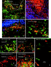
In situ phenotypic characterization of CD123high P-DCs and related MxA and PNAd expression. Multicolor immunofluorescence staining for: CD123 (Cy3, red), CD45RA (FITC, green), and cytokeratin (AMCA, blue in c) (a, c, and d); human MxA (Cy3, red) with nuclear counterstain (DAPI, blue) (b); CD123 (Cy3, red) (e–g) and HLA-DR (FITC, green) (e), CD68 (FITC, green) (f), human c-kit (Alexa Fluor 488, green) (g); CD3 (Cy3, red) and CD45RA (FITC, green) (h); and PNAd revealed with mAb MECA-79 (Cy3, red) and endothelium with Ulex europaeus lectin-1 (FITC, green) (i) in serial sections (comparable fields in a and b) of cutaneous DLE lesion. Characteristic accumulation of CD123highCD45RA+ cells (yellow staining) along the dermal-epidermal junction (a), around hair follicles (c), and adjacent to dermal vessel (asterisk) with endothelial CD123 expression (d); note strong expression of MxA in keratinocytes and infiltrating cells in dermis (b) in field comparable to high numbers of P-DCs (a). CD123high cells also express HLA-DR (e) and CD68 (f); the intracellular dot-like staining pattern for CD68 was similar in tonsillar tissue (not shown) and found characteristic for DCs by others. Dermal c-kit+ mast cells do not express CD123 (g, arrowhead); note the large number of naïve (CD3+CD45RA+) T cells (yellow staining, some arrowed) (h). Medium–sized vessels (diameter, ≥10 μm) with strong PNAd expression appears yellow (i); note that some keratinocytes also react with MECA as previously noted. Basement membrane of epidermis is indicated by dashed line. Original magnifications: ×200 (a and b); ×400 (c, d, g, and h); ×630 (e and f); ×100 (i).
Figure 2.
Relationship between the density (cell/mm2) of P-DCs (CD123highCD45RA+ cells) and MxA+ cells in cutaneous LE lesions (r _s_= 0.79, P < 0.0005, n = 15). Based on data in Table 1 ▶ and Spearman’s rank correlation test.
Similar articles
- Experimentally induced recruitment of plasmacytoid (CD123high) dendritic cells in human nasal allergy.
Jahnsen FL, Lund-Johansen F, Dunne JF, Farkas L, Haye R, Brandtzaeg P. Jahnsen FL, et al. J Immunol. 2000 Oct 1;165(7):4062-8. doi: 10.4049/jimmunol.165.7.4062. J Immunol. 2000. PMID: 11034417 - Involvement of plasmacytoid dendritic cells in human diseases.
Jahnsen FL, Farkas L, Lund-Johansen F, Brandtzaeg P. Jahnsen FL, et al. Hum Immunol. 2002 Dec;63(12):1201-5. doi: 10.1016/s0198-8859(02)00759-0. Hum Immunol. 2002. PMID: 12480264 Review. - Enhanced type I interferon signalling promotes Th1-biased inflammation in cutaneous lupus erythematosus.
Wenzel J, Wörenkämper E, Freutel S, Henze S, Haller O, Bieber T, Tüting T. Wenzel J, et al. J Pathol. 2005 Mar;205(4):435-42. doi: 10.1002/path.1721. J Pathol. 2005. PMID: 15685590 - Interferon-α in the generation of monocyte-derived dendritic cells: recent advances and implications for dermatology.
Farkas A, Kemény L. Farkas A, et al. Br J Dermatol. 2011 Aug;165(2):247-54. doi: 10.1111/j.1365-2133.2011.10301.x. Epub 2011 Jun 2. Br J Dermatol. 2011. PMID: 21410666 Review. - The expression pattern of interferon-inducible proteins reflects the characteristic histological distribution of infiltrating immune cells in different cutaneous lupus erythematosus subsets.
Wenzel J, Zahn S, Mikus S, Wiechert A, Bieber T, Tüting T. Wenzel J, et al. Br J Dermatol. 2007 Oct;157(4):752-7. doi: 10.1111/j.1365-2133.2007.08137.x. Epub 2007 Aug 21. Br J Dermatol. 2007. PMID: 17714558
Cited by
- Therapeutically targeting proinflammatory type I interferons in systemic lupus erythematosus: efficacy and insufficiency with a specific focus on lupus nephritis.
Lai B, Luo SF, Lai JH. Lai B, et al. Front Immunol. 2024 Oct 16;15:1489205. doi: 10.3389/fimmu.2024.1489205. eCollection 2024. Front Immunol. 2024. PMID: 39478861 Free PMC article. Review. - Population pharmacokinetics of sifalimumab, an investigational anti-interferon-α monoclonal antibody, in systemic lupus erythematosus.
Narwal R, Roskos LK, Robbie GJ. Narwal R, et al. Clin Pharmacokinet. 2013 Nov;52(11):1017-27. doi: 10.1007/s40262-013-0085-2. Clin Pharmacokinet. 2013. PMID: 23754736 Free PMC article. Clinical Trial. - Value of CD123 Immunohistochemistry and Elastic Staining in Differentiating Discoid Lupus Erythematosus from Lichen Planopilaris.
Aslani FS, Sepaskhah M, Bagheri Z, Akbarzadeh-Jahromi M. Aslani FS, et al. Int J Trichology. 2020 Mar-Apr;12(2):62-67. doi: 10.4103/ijt.ijt_32_20. Epub 2020 May 5. Int J Trichology. 2020. PMID: 32684677 Free PMC article. - Rheumatoid arthritis synovium contains plasmacytoid dendritic cells.
Cavanagh LL, Boyce A, Smith L, Padmanabha J, Filgueira L, Pietschmann P, Thomas R. Cavanagh LL, et al. Arthritis Res Ther. 2005;7(2):R230-40. doi: 10.1186/ar1467. Epub 2005 Jan 11. Arthritis Res Ther. 2005. PMID: 15743469 Free PMC article. - Interferon pathway in SLE: one key to unlocking the mystery of the disease.
Rönnblom L, Leonard D. Rönnblom L, et al. Lupus Sci Med. 2019 Aug 13;6(1):e000270. doi: 10.1136/lupus-2018-000270. eCollection 2019. Lupus Sci Med. 2019. PMID: 31497305 Free PMC article. Review.
References
- Cella M, Jarrossay D, Facchetti F, Alebardi O, Nakajima H, Lanzavecchia A, Colonna M: Plasmacytoid monocytes migrate to inflamed lymph nodes and produce large amounts of type I interferon. Nat Med 1999, 5:919-923 - PubMed
- Strobl H, Scheinecker C, Riedl E, Csmarits B, Bello-Fernandez C, Pickl WF, Majdic O, Knapp W: Identification of CD68+lin− peripheral blood cells with dendritic precursor characteristics. J Immunol 1998, 161:740-748 - PubMed
- Jahnsen F, Lund-Johansen F, Dunne J, Farkas L, Haye R, Brandtzaeg P: Experimentally induced recruitment of plasmacytoid (CD123high) dendritic cells in human nasal allergy. J Immunol 2000, 165:4062-4068 - PubMed
Publication types
MeSH terms
Substances
LinkOut - more resources
Full Text Sources
Other Literature Sources
Medical
Research Materials

