Circulating insulin-like growth factor I mediates the protective effects of physical exercise against brain insults of different etiology and anatomy - PubMed (original) (raw)
Comparative Study
Circulating insulin-like growth factor I mediates the protective effects of physical exercise against brain insults of different etiology and anatomy
E Carro et al. J Neurosci. 2001.
Abstract
Physical exercise ameliorates age-related neuronal loss and is currently recommended as a therapeutical aid in several neurodegenerative diseases. However, evidence is still lacking to firmly establish whether exercise constitutes a practical neuroprotective strategy. We now show that exercise provides a remarkable protection against brain insults of different etiology and anatomy. Laboratory rodents were submitted to treadmill running (1 km/d) either before or after neurotoxin insult of the hippocampus (domoic acid) or the brainstem (3-acetylpyridine) or along progression of inherited neurodegeneration affecting the cerebellum (Purkinje cell degeneration). In all cases, animals show recovery of behavioral performance compared with sedentary ones, i.e., intact spatial memory in hippocampal-injured mice, and normal or near to normal motor coordination in brainstem- and cerebellum-damaged animals. Furthermore, exercise blocked neuronal impairment or loss in all types of injuries. Because circulating insulin-like growth factor I (IGF-I), a potent neurotrophic hormone, mediates many of the effects of exercise on the brain, we determined whether neuroprotection by exercise is mediated by IGF-I. Indeed, subcutaneous administration of a blocking anti-IGF-I antibody to exercising animals to inhibit exercise-induced brain uptake of IGF-I abrogates the protective effects of exercise in all types of lesions; antibody-treated animals showed sedentary-like brain damage. These results indicate that exercise prevents and protects from brain damage through increased uptake of circulating IGF-I by the brain. The practice of physical exercise is thus strongly recommended as a preventive measure against neuronal demise. These findings also support the use of IGF-I as a therapeutical aid in brain diseases coursing with either acute or progressive neuronal death.
Figures
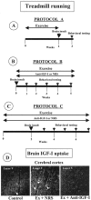
Fig. 1.
A–C, Treadmill running and anti-IGF-I delivery schedules. A, Protocol A: exercise before brain insult. Animals run during 15 d before brain insult (3AP in rats and domoic acid in mice). Behavioral testing was conducted 5–7 d later. B, Protocol B: exercise after brain insult. Brain-damaged animals ran for 4–5 weeks and were evaluated in the rotarod once per week (pcd mice and 3AP-injected rats).C, Protocol C: animals exercised both before and after brain insult (3AP). Behavioral testing was also done every week. In a parallel series of experiments, an anti-IGF-I infusion was delivered in protocols B and C to exercising animals. Control exercising animals received an NRS infusion. In all protocols, animals were killed for anatomical evaluation after the last behavioral evaluation.D, IGF-I antiserum inhibits exercise-induced brain accumulation. Control, Sedentary animals show negligible IGF-I immunostaining in the brain, whereas exercised animals receiving NRS (Ex + NRS) show a marked increase that is inhibited when an anti-IGF-I infusion is administered simultaneously (Ex + Anti-IGF-I). A representative brain area is shown.
Fig. 2.
Exercise prevents behavioral deficits after brain damage. A, Rats undergoing exercise training before 3AP injection, although motor-impaired compared with control animals (*p < 0.005), show significantly better motor coordination in the rotarod than sedentary 3AP rats (*p < 0.009). B, C, Mice submitted to treadmill running before injection of domoic acid have intact learning (B) and memory (C) performance, whereas sedentary mice have significantly impaired acquisition and retention scores (*p < 0.0001). Error bars are smaller than the size of the symbols at some points.
Fig. 3.
Exercise induces recovery of behavioral performance in ongoing neurodegeneration. A, Rats submitted to treadmill running after 3AP insult gradually recover motor coordination and reach normal performance after 5 weeks of running (*p < 0.002 vs 3AP). B, pcd mice with moderate, albeit significantly impaired motor coordination underwent exercise training and recovered normal motor performance within 1 week. They kept normal motor coordination for the duration of the study, whereas sedentary pcd mice remained ataxic. However, exercising pcd mice simultaneously receiving an anti-IGF-I infusion did not recover limb coordination (*p < 0.001).C, Rats were submitted both before and after 3AP insult to treadmill running with simultaneous infusion of NRS and recovered full motor coordination after 5 weeks. However, rats receiving simultaneously an anti-IGF-I infusion remained severely impaired [*p < 0.01 vs 3AP and _(Ex + 3AP + Ex) + Anti-IGF-I_].
Fig. 4.
Exercise prevents neuronal loss and impairment in an IGF-I dependent manner. A, 3AP-injected rats showed profound neuronal impairment as determined by a drastic decrease in calbindin-positive cells in the IO. Exercised animals showed only a moderate, nonsignificant decrease in the number of calbindin-positive IO neurons. However, exercising animals simultaneously receiving an anti-IGF-I infusion showed sedentary-like neuronal impairment.Inset, Representative brainstem sections of the different experimental groups showing calbindin staining in the IO. Note the marked absence of calbindin-positive cell bodies in brainstem sections of sedentary 3AP and exercised plus anti-IGF-I 3AP rats. *p < 0.01. B, Domoic acid-injured mice show full protection against neuronal loss by exercise. Again, anti-IGF-I administration obliterated the protective effects of exercise. Inset, Representative Nissl-stained sections of corresponding experimental groups. Loss of neurons after domoic acid was assessed in the hippocampal hilus (Hil) by counting Nissl-stained cells. *p < 0.02 and **p < 0.01 versus respective controls.C, Sedentary pcd mice show a profound loss of calbindin staining of Purkinje cells in the cerebellar cortex. Exercising pcd mice show normal numbers of calbindin-positive Purkinje cells, whereas exercised plus anti-IGF-I-treated pcd mice have significantly reduced numbers of calbindin-positive Purkinje cells, similar to sedentary pcd mice. Inset, Representative cerebellar cortex sections of the different experimental groups. Note the marked depletion of calbindin-positive cells in the Purkinje cell layer (PC). ML, Molecular layer of the cerebellum; GL, granule cell layer. **p < 0.0001 versus respective control group.
Fig. 5.
Exercise and IGF-I increase brain glucose uptake in brain-damaged animals. Domoic acid-lesioned mice (b) show increased glucose uptake in the hippocampus compared with nonlesioned mice (a). Glucose uptake is further increased by either exercise (d) or IGF-I (c) not only in the hippocampus but also in other telencephalic areas. Representative brain autoradiography hemisections are shown.CA2, CA3, Hippocampal pyramidal cell layers; DG, hippocampal dentate gyrus; V, ventral; D, dorsal.
Similar articles
- Neuroprotective actions of peripherally administered insulin-like growth factor I in the injured olivo-cerebellar pathway.
Fernandez AM, Gonzalez de la Vega AG, Planas B, Torres-Aleman I. Fernandez AM, et al. Eur J Neurosci. 1999 Jun;11(6):2019-30. doi: 10.1046/j.1460-9568.1999.00623.x. Eur J Neurosci. 1999. PMID: 10336671 - Circulating insulin-like growth factor I mediates exercise-induced increases in the number of new neurons in the adult hippocampus.
Trejo JL, Carro E, Torres-Aleman I. Trejo JL, et al. J Neurosci. 2001 Mar 1;21(5):1628-34. doi: 10.1523/JNEUROSCI.21-05-01628.2001. J Neurosci. 2001. PMID: 11222653 Free PMC article. - Astrocyte-Specific Overexpression of Insulin-Like Growth Factor-1 Protects Hippocampal Neurons and Reduces Behavioral Deficits following Traumatic Brain Injury in Mice.
Madathil SK, Carlson SW, Brelsfoard JM, Ye P, D'Ercole AJ, Saatman KE. Madathil SK, et al. PLoS One. 2013 Jun 27;8(6):e67204. doi: 10.1371/journal.pone.0067204. Print 2013. PLoS One. 2013. PMID: 23826235 Free PMC article. - Role of insulin-like growth factor I signaling in neurodegenerative diseases.
Trejo JL, Carro E, Garcia-Galloway E, Torres-Aleman I. Trejo JL, et al. J Mol Med (Berl). 2004 Mar;82(3):156-62. doi: 10.1007/s00109-003-0499-7. Epub 2003 Nov 28. J Mol Med (Berl). 2004. PMID: 14647921 Review. - Glycosaminoglycans co-administration enhance insulin-like growth factor-I neuroprotective and neuroregenerative activity in traumatic and genetic models of motor neuron disease: a review.
Di Giulio AM, Germani E, Lesma E, Muller E, Gorio A. Di Giulio AM, et al. Int J Dev Neurosci. 2000 Jul-Aug;18(4-5):339-46. doi: 10.1016/s0736-5748(00)00015-0. Int J Dev Neurosci. 2000. PMID: 10817918 Review.
Cited by
- Efficacy of exercise rehabilitation for managing patients with Alzheimer's disease.
Li D, Jia J, Zeng H, Zhong X, Chen H, Yi C. Li D, et al. Neural Regen Res. 2024 Oct 1;19(10):2175-2188. doi: 10.4103/1673-5374.391308. Epub 2023 Dec 21. Neural Regen Res. 2024. PMID: 38488551 Free PMC article. - The Role of Insulin-like Growth Factor I in Mechanisms of Resilience and Vulnerability to Sporadic Alzheimer's Disease.
Zegarra-Valdivia JA, Pignatelli J, Nuñez A, Torres Aleman I. Zegarra-Valdivia JA, et al. Int J Mol Sci. 2023 Nov 17;24(22):16440. doi: 10.3390/ijms242216440. Int J Mol Sci. 2023. PMID: 38003628 Free PMC article. Review. - A framework of transient hypercapnia to achieve an increased cerebral blood flow induced by nasal breathing during aerobic exercise.
Moris JM, Cardona A, Hinckley B, Mendez A, Blades A, Paidisetty VK, Chang CJ, Curtis R, Allen K, Koh Y. Moris JM, et al. Cereb Circ Cogn Behav. 2023 Sep 13;5:100183. doi: 10.1016/j.cccb.2023.100183. eCollection 2023. Cereb Circ Cogn Behav. 2023. PMID: 37745894 Free PMC article. Review. - A New, Simple and Practical Approach to Increase the Effects of Aerobic Exercise on Serum Levels of Neurotrophic Factors in Adult Males.
Bahramnejad M, Dehnou VV, Eslami R. Bahramnejad M, et al. Int J Exerc Sci. 2023 Aug 1;16(2):932-941. eCollection 2023. Int J Exerc Sci. 2023. PMID: 37650037 Free PMC article. - A review of combined neuromodulation and physical therapy interventions for enhanced neurorehabilitation.
Evancho A, Tyler WJ, McGregor K. Evancho A, et al. Front Hum Neurosci. 2023 Jul 21;17:1151218. doi: 10.3389/fnhum.2023.1151218. eCollection 2023. Front Hum Neurosci. 2023. PMID: 37545593 Free PMC article. Review.
References
- Amaducci L, Tesco G. Aging as a major risk for degenerative diseases of the central nervous system. Curr Opin Neurol. 1994;7:283–286. - PubMed
- Arkin SM. Elder rehab: a student-supervised exercise program for Alzheimer's patients. Gerontologist. 1999;39:729–735. - PubMed
- Azcoitia I, Sierra A, Veiga S, Honda S, Harada N, Garcia-Segura LM. Brain aromatase is neuroprotective. J Neurobiol. 2001;47:318–329. - PubMed
- Beal MF. Energetics in the pathogenesis of neurodegenerative diseases. Trends Neurosci. 2000;23:298–304. - PubMed
Publication types
MeSH terms
Substances
LinkOut - more resources
Full Text Sources
Medical


