Mitosis, microtubules, and the matrix - PubMed (original) (raw)
Review
Mitosis, microtubules, and the matrix
J M Scholey et al. J Cell Biol. 2001.
Abstract
The mechanical events of mitosis depend on the action of microtubules and mitotic motors, but whether these spindle components act alone or in concert with a spindle matrix is an important question.
Figures
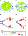
Figure 1.
Skeletor and the microtrabecular lattice. (A and B) The skeletor matrix. Skeletor forms a reticular matrix around condensing chromosomes in the absence of MTs during prophase (A). As the nuclear envelope fenestrates and microtubules enter the nuclear region during prometaphase, the skeletor matrix organizes and stabilizes microtubules in the central spindle, providing support for the fusiform morphology of the spindle through metaphase (B). (C and D) The microtrabecular lattice matrix. A spring-like lattice associated with chromosomes forms around kinetochore microtubules. During prometaphase and metaphase (C) this lattice is deformed or stretched toward the metaphase plate by plus end–directed motors (shown attached to the fibrous corona). At anaphase (D), these motors are turned off and the elastic recoil of the matrix drives the poleward movement of chromosomes.
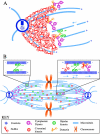
Figure 2.
The NuMA matrix and MT–MT cross-linking motors. (A) The NuMA matrix. The NuMA protein oligomerizes into a highly branched and cross-linked lattice around the spindle poles. Because NuMA is believed to associate with both microtubules and certain mitotic motors, such a matrix could anchor and cross-link microtubules and also immobilize motors at or near the poles. The latter activity would provide a stationary substrate for motor-driven MT transport within the spindle, allowing minus end–directed motors such as cytoplasmic dynein to focus the minus ends of MTs at the poles and plus end–directed motors such as bipolar kinesins to cross-link polar microtubules into asters and drive the poleward flux of kinetochore microtubules. (B) A matrix of MT–MT cross-linking and sliding motors. The spindle is packed with a dense array of MT–MT cross-linking and sliding motors. Specific interactions that occur between these motors and spindle MTs drive the formation and function of the spindle. (Inset, top left) Bipolar kinesins such as KLP61F can cross-link parallel MTs into bundles, thus contributing to the organization of MTs in the half spindles, but generate no net axial force between them. (Inset, top right) In contrast, when bipolar kinesins cross-link antiparallel MTs into bundles, they can generate paraxial force and thus slide them in relation to one another. Asymmetric motors like dynein and Ncd can presumably cross-link and slide either parallel or antiparallel MTs in relation to one another, dependent upon the nature of the binding between their nucleotide-insensitive MT binding site and the MT surface lattice, and the polarity of motion driven by their motor domains.
Similar articles
- hNuf2 inhibition blocks stable kinetochore-microtubule attachment and induces mitotic cell death in HeLa cells.
DeLuca JG, Moree B, Hickey JM, Kilmartin JV, Salmon ED. DeLuca JG, et al. J Cell Biol. 2002 Nov 25;159(4):549-55. doi: 10.1083/jcb.200208159. Epub 2002 Nov 18. J Cell Biol. 2002. PMID: 12438418 Free PMC article. - Titin in insect spermatocyte spindle fibers associates with microtubules, actin, myosin and the matrix proteins skeletor, megator and chromator.
Fabian L, Xia X, Venkitaramani DV, Johansen KM, Johansen J, Andrew DJ, Forer A. Fabian L, et al. J Cell Sci. 2007 Jul 1;120(Pt 13):2190-204. doi: 10.1242/jcs.03465. J Cell Sci. 2007. PMID: 17591688 - Ska1 cooperates with DDA3 for spindle dynamics and spindle attachment to kinetochore.
Park JE, Song H, Kwon HJ, Jang CY. Park JE, et al. Biochem Biophys Res Commun. 2016 Feb 12;470(3):586-592. doi: 10.1016/j.bbrc.2016.01.101. Epub 2016 Jan 19. Biochem Biophys Res Commun. 2016. PMID: 26797278 - Unconventional Roles of Cytoskeletal Mitotic Machinery in Neurodevelopment.
Del Castillo U, Norkett R, Gelfand VI. Del Castillo U, et al. Trends Cell Biol. 2019 Nov;29(11):901-911. doi: 10.1016/j.tcb.2019.08.006. Epub 2019 Oct 6. Trends Cell Biol. 2019. PMID: 31597609 Free PMC article. Review. - A guide to classifying mitotic stages and mitotic defects in fixed cells.
Baudoin NC, Cimini D. Baudoin NC, et al. Chromosoma. 2018 Jun;127(2):215-227. doi: 10.1007/s00412-018-0660-2. Epub 2018 Feb 6. Chromosoma. 2018. PMID: 29411093 Review.
Cited by
- The molecular basis of anaphase A in animal cells.
Rath U, Sharp DJ. Rath U, et al. Chromosome Res. 2011 Apr;19(3):423-32. doi: 10.1007/s10577-011-9199-2. Chromosome Res. 2011. PMID: 21461696 - Altering membrane topology with Sar1 does not impair spindle assembly in Xenopus egg extracts.
Riggs B, Bergman ZJ, Heald R. Riggs B, et al. Cytoskeleton (Hoboken). 2012 Aug;69(8):591-9. doi: 10.1002/cm.21036. Epub 2012 May 17. Cytoskeleton (Hoboken). 2012. PMID: 22605651 Free PMC article. - Directly probing the mechanical properties of the spindle and its matrix.
Gatlin JC, Matov A, Danuser G, Mitchison TJ, Salmon ED. Gatlin JC, et al. J Cell Biol. 2010 Feb 22;188(4):481-9. doi: 10.1083/jcb.200907110. J Cell Biol. 2010. PMID: 20176922 Free PMC article. - Model of chromosome motility in Drosophila embryos: adaptation of a general mechanism for rapid mitosis.
Civelekoglu-Scholey G, Sharp DJ, Mogilner A, Scholey JM. Civelekoglu-Scholey G, et al. Biophys J. 2006 Jun 1;90(11):3966-82. doi: 10.1529/biophysj.105.078691. Epub 2006 Mar 13. Biophys J. 2006. PMID: 16533843 Free PMC article. - Early spindle assembly in Drosophila embryos: role of a force balance involving cytoskeletal dynamics and nuclear mechanics.
Cytrynbaum EN, Sommi P, Brust-Mascher I, Scholey JM, Mogilner A. Cytrynbaum EN, et al. Mol Biol Cell. 2005 Oct;16(10):4967-81. doi: 10.1091/mbc.e05-02-0154. Epub 2005 Aug 3. Mol Biol Cell. 2005. PMID: 16079179 Free PMC article.
References
- Blangy, A., L. Arnaud, and E.A. Nigg. 1997. Phosphorylation by p34cdc2 protein kinase regulates binding of the kinesin-related motor HsEg5 to the dynactin subunit p150Glued. J. Biol. Chem. 272:19418–19424. - PubMed
- Compton, D.A. 2000. Spindle assembly in animal cells. Annu. Rev. Biochem. 69:95–114. - PubMed
- Cullen, C.F., and H. Ohkura. 2001. Msps protein is localized to acentrosomal poles to ensure bipolarity of Drosophila meiotic spindles. Nat. Cell Biol. 3:637–642. - PubMed
Publication types
MeSH terms
Substances
LinkOut - more resources
Full Text Sources
Molecular Biology Databases

