Regulation of cyclin D2 gene expression by the Myc/Max/Mad network: Myc-dependent TRRAP recruitment and histone acetylation at the cyclin D2 promoter - PubMed (original) (raw)
Regulation of cyclin D2 gene expression by the Myc/Max/Mad network: Myc-dependent TRRAP recruitment and histone acetylation at the cyclin D2 promoter
C Bouchard et al. Genes Dev. 2001.
Abstract
Myc oncoproteins promote cell cycle progression in part through the transcriptional up-regulation of the cyclin D2 gene. We now show that Myc is bound to the cyclin D2 promoter in vivo. Binding of Myc induces cyclin D2 expression and histone acetylation at a single nucleosome in a MycBoxII/TRRAP-dependent manner. Down-regulation of cyclin D2 mRNA expression in differentiating HL60 cells is preceded by a switch of promoter occupancy from Myc/Max to Mad/Max complexes, loss of TRRAP binding, increased HDAC1 binding, and histone deacetylation. Thus, recruitment of TRRAP and regulation of histone acetylation are critical for transcriptional activation by Myc.
Figures
Figure 1
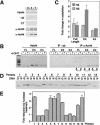
In vivo binding of MycER proteins at the mouse cyclin D2 promoter. (A) Schematic drawing of the mouse cyclin D2 promoter from −1100 bp to the transcriptional start site (+1). (Black boxes) Two E-boxes (E3 and E4); (arrows) primer pairs (FL, E3, and E4); (thick lines) primer sets used for mapping; (hatched bars) position of nucleosomes. (B) ChIP assays using α-Myc antibodies or antibodies directed against the hormone-binding domain of the estrogen receptor (ER). As control, either no antibody or irrelevant control antibody (α-p27) were used. Immunoprecipitated DNA was analyzed by PCR using the indicated primer pairs. Inputs and genomic DNA (gen. DNA) are shown as control. (C) Quantitation of MycER binding to the cyclin D2 promoter after addition of hormone. MycER cells were serum-starved for 72 h and either left untreated or treated for 1 or 6 h with 250 nM 4-OHT, respectively. Subsequently, ChIP assays were conducted as described in (B). The signals obtained in the PCR were quantified and plotted relative to the signal obtained from −ab precipitation. (4-OHT) 4-hydroxytamoxifen.
Figure 2
Myc-regulated histone acetylation at the cyclin D2 promoter. (A) Immunoprecipitation of chromatin from serum-starved MycER cells either left untreated (0) or treated for 6 h with 250 nM 4-OHT (4) or 100 ng/mL trichostatin A (T) using anti-acetylated histones H3 (AcH3) or H4 (AcH4) antibodies. As control, either no antibody (−ab) or irrelevant control antibody (α-p27, labeled CT) was used. Precipitated chromatin was PCR-amplified using FL primers. (B) Anti-AcH4 immunoprecipitation (IP) of chromatin isolated from serum-starved MycER cells either left untreated (−) or treated for 6 h (+) with 250 nM 4-OHT and analyzed by PCR using three different primer pairs FL, E3, and E4 (depicted in Fig. 1A). (C) Quantitation of the average changes of histone acetylation observed in several independent experiments. (D) Mapping of nucleosomal positions in the cyclin D2 promoter. Chromatin was incubated with micrococcal nuclease to digest internucleosomal DNA. Nucleosomal DNA then was purified. Parallel PCRs were conducted with the primer pairs depicted in Fig. 1A from either undigested (G) or nucleosomal DNA (N) and analyzed on a polyacrylamide gel. (E) Quantitative representation of C documenting the fold reduction in signal intensity by nuclease treatment for each primer pair.
Figure 3
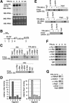
Essential role of MycBoxII in activation of mouse cyclin D2 expression. (A) Schematic representation of wild-type Myc (WTMyc) and the MycBoxII (MBII) deletion mutant (ΔMBIIMyc) used in this study. (NLS) Nuclear localization signal; (HLH) helix–loop–helix; (LZ) leucine zipper. (B) (left) Western blot with α-ER antibodies documenting expression levels of MycER proteins in WTMycER and ΔMBIIMycER clones (designated #1 and #2). (right) Northern blots documenting expression of cyclin D2 and GAPDH mRNAs in WTMycER and ΔMBIIMycER#1 clones before (−) and 6 h after (+) addition of 250 nM 4-OHT. (WT) wild type; (WB) Western blot. (C) ChIP assays using either α-AcH3, α-AcH4, or control antibodies from serum-starved WTMycER and ΔMBIIMycER#1 clones either left untreated (0) or incubated for 6 h with 250 nM 4-OHT (4) or 100 ng/mL trichostatin A (T). Precipitated DNA was analyzed by PCR with primer pairs spanning either both E-boxes (FL) or the E3 E-box of the cyclin D2 promoter. (CT) control. (D) ChIP assays (performed as described in C) from a clone expressing high levels of ΔMBIIMycER (clone #2, see B). (E) ChIP assays with the indicated antibodies from serum-starved MycER cells either left untreated or treated for 6 h with 250 nM 4-OHT. PCRs were performed with primer pairs specific for the E3 E-box. (F) Quantitation of PCR assays performed with the E3 primers and chromatin immunoprecipitated from WTMycER and ΔMBIIMycER#1 clones before (−) and after (+) addition of 250 nM 4-OHT.
Figure 4
Interaction of endogenous Myc/Max/Mad with the cyclin D2 promoter and regulation of histone acetylation in HL60 cells. (A) Northern blot documenting cyclin D2 expression on treatment of HL60 with TPA for the indicated times. The ethidium bromide-stained gel is displayed as control. (B) Schematic drawing of the human cyclin D2 locus. (Black boxes) The two E-boxes located within the first 2.5 kb of the cyclin D2 promoter. (Black bars) Location of the amplified fragments. (UTR) untranslated region. (C) ChIP assays documenting interaction of endogenous Myc, Max, and Mad1 proteins with the cyclin D2 promoter. HL60 cells were treated as in A, and ChIP assays were conducted with the indicated antibodies; precipitates were analyzed using primers specific for fragment 2 (top). Primers specific for a chromosome 22 E box were used as control (bottom). (exp.) Exponentially growing cells. (D) Quantitation of Myc binding in exponentially growing HL60 cells and of Mad1 binding in TPA differentiated HL60 cells to the five fragments of the cyclin D2 promoter as depicted in (B). Relative DNA binding defines the signal intensities obtained from the ChIP assays normalized to the input signals. (E) ChIP assays using either α-AcH3 or α-AcH4 antibodies of either untreated or TPA-treated (48 h) HL60 cells. Primers specific for fragment 2 of human cyclin D2 (top), a 5S rRNA locus (middle), and chromosome 22 E-box (bottom) were used. The input lanes show a dilution series in fourfold steps documenting the linearity of the assays. (F) Time course experiment documenting changes in promoter occupancy and histone acetylation on treatment of HL60 cells with TPA for the indicated time periods (fragment 2). Pol II occupancy was determined with fragment 4. (G) Regulated binding of TRRAP and HDAC1 to the cyclin D2 promoter. The experimental setup was as above.
Similar articles
- Mnt transcriptional repressor is functionally regulated during cell cycle progression.
Popov N, Wahlström T, Hurlin PJ, Henriksson M. Popov N, et al. Oncogene. 2005 Dec 15;24(56):8326-37. doi: 10.1038/sj.onc.1208961. Oncogene. 2005. PMID: 16103876 - Down-regulation of TRRAP-dependent hTERT and TRRAP-independent CAD activation by Myc/Max contributes to the differentiation of HL60 cells after exposure to DMSO.
Jiang G, Bi K, Tang T, Wang J, Zhang Y, Zhang W, Ren H, Bai H, Wang Y. Jiang G, et al. Int Immunopharmacol. 2006 Jul;6(7):1204-13. doi: 10.1016/j.intimp.2006.02.014. Epub 2006 Apr 7. Int Immunopharmacol. 2006. PMID: 16714225 - Direct induction of cyclin D2 by Myc contributes to cell cycle progression and sequestration of p27.
Bouchard C, Thieke K, Maier A, Saffrich R, Hanley-Hyde J, Ansorge W, Reed S, Sicinski P, Bartek J, Eilers M. Bouchard C, et al. EMBO J. 1999 Oct 1;18(19):5321-33. doi: 10.1093/emboj/18.19.5321. EMBO J. 1999. PMID: 10508165 Free PMC article. - The basic region/helix-loop-helix/leucine zipper domain of Myc proto-oncoproteins: function and regulation.
Lüscher B, Larsson LG. Lüscher B, et al. Oncogene. 1999 May 13;18(19):2955-66. doi: 10.1038/sj.onc.1202750. Oncogene. 1999. PMID: 10378692 Review. - Repression by the Mad(Mxi1)-Sin3 complex.
Schreiber-Agus N, DePinho RA. Schreiber-Agus N, et al. Bioessays. 1998 Oct;20(10):808-18. doi: 10.1002/(SICI)1521-1878(199810)20:10<808::AID-BIES6>3.0.CO;2-U. Bioessays. 1998. PMID: 9819568 Review.
Cited by
- Myocardial Mycn is essential for mouse ventricular wall morphogenesis.
Harmelink C, Peng Y, DeBenedittis P, Chen H, Shou W, Jiao K. Harmelink C, et al. Dev Biol. 2013 Jan 1;373(1):53-63. doi: 10.1016/j.ydbio.2012.10.005. Epub 2012 Oct 12. Dev Biol. 2013. PMID: 23063798 Free PMC article. - TRRAP-dependent and TRRAP-independent transcriptional activation by Myc family oncoproteins.
Nikiforov MA, Chandriani S, Park J, Kotenko I, Matheos D, Johnsson A, McMahon SB, Cole MD. Nikiforov MA, et al. Mol Cell Biol. 2002 Jul;22(14):5054-63. doi: 10.1128/MCB.22.14.5054-5063.2002. Mol Cell Biol. 2002. PMID: 12077335 Free PMC article. - Novel cytological model for the identification of early oral cancer diagnostic markers: The carcinoma sequence model.
Kawaharada M, Yamazaki M, Maruyama S, AbÉ T, Chan NN, Kitano T, Kobayashi T, Maeda T, Tanuma JI. Kawaharada M, et al. Oncol Lett. 2022 Mar;23(3):76. doi: 10.3892/ol.2022.13196. Epub 2022 Jan 11. Oncol Lett. 2022. PMID: 35111245 Free PMC article. - Dual regulation of c-Myc by p300 via acetylation-dependent control of Myc protein turnover and coactivation of Myc-induced transcription.
Faiola F, Liu X, Lo S, Pan S, Zhang K, Lymar E, Farina A, Martinez E. Faiola F, et al. Mol Cell Biol. 2005 Dec;25(23):10220-34. doi: 10.1128/MCB.25.23.10220-10234.2005. Mol Cell Biol. 2005. PMID: 16287840 Free PMC article. - The TRRAP transcription cofactor represses interferon-stimulated genes in colorectal cancer cells.
Detilleux D, Raynaud P, Pradet-Balade B, Helmlinger D. Detilleux D, et al. Elife. 2022 Mar 4;11:e69705. doi: 10.7554/eLife.69705. Elife. 2022. PMID: 35244540 Free PMC article.
References
- Alland L, Muhle R, Hou H, Jr, Potes J, Chin L, Schreiber-Agus N, DePinho RA. Role for N-CoR and histone deacetylase in Sin3-mediated transcriptional repression. Nature. 1997;387:49–55. - PubMed
- Amati B, Frank SR, Donjerkovic D, Taubert S. Function of the c-Myc oncoprotein in chromatin remodeling and transcription. Biochim Biophys Acta. 2001;1471:M135–M145. - PubMed
- Ayer DE, Eisenman RN. A switch from Myc:Max to Mad:Max heterocomplexes accompanies monocyte/macrophage differentiation. Genes & Dev. 1993;7:2110–2119. - PubMed
Publication types
MeSH terms
Substances
LinkOut - more resources
Full Text Sources
Other Literature Sources
Miscellaneous


