Interaction of classical swine fever virus with membrane-associated heparan sulfate: role for virus replication in vivo and virulence - PubMed (original) (raw)
Comparative Study
Interaction of classical swine fever virus with membrane-associated heparan sulfate: role for virus replication in vivo and virulence
M M Hulst et al. J Virol. 2001 Oct.
Abstract
Passage of native classical swine fever virus (CSFV) in cultured swine kidney cells (SK6 cells) selects virus variants that attach to the surface of cells by interaction with membrane-associated heparan sulfate (HS). A Ser-to-Arg change in the C terminus of envelope glycoprotein E(rns) (amino acid 476 in the open reading frame of CSFV) is responsible for selection of these HS-binding virus variants (M. M. Hulst, H. G. P. van Gennip, and R. J. M. Moormann, J. Virol. 74:9553-9561, 2000). In this investigation we studied the role of binding of CSFV to HS in vivo. Using reverse genetics, an HS-independent recombinant virus (S-ST virus) with Ser(476) and an HS-dependent recombinant virus (S-RT virus) with Arg(476) were constructed. Animal experiments indicated that this adaptive Ser-to-Arg mutation had no effect on the virulence of CSFV. Analysis of viruses reisolated from pigs infected with these recombinant viruses indicated that replication in vivo introduced no mutations in the genes of the envelope proteins E(rns), E1, and E2. However, the blood of one of the three pigs infected with the S-RT virus contained also a low level of virus particles that, when grown under a methylcellulose overlay, produced relative large plaques, characteristic of an HS-independent virus. Sequence analysis of such a large-plaque phenotype showed that Arg(476) was mutated back to Ser(476). Removal of HS from the cell surface and addition of heparin to the medium inhibited infection of cultured (SK6) and primary swine kidney cells with S-ST virus reisolated from pigs by about 70% whereas infection with the administered S-ST recombinant virus produced in SK6 cells was not affected. Furthermore, E(rns) S-ST protein, produced in insect cells, could bind to immobilized heparin and to HS chains on the surface of SK6 cells. These results indicated that S-ST virus generated in pigs is able to infect cells by an HS-dependent mechanism. Binding of concanavalin A (ConA) to virus particles stimulated the infection of SK6 cells with S-ST virus produced in these cells by 12-fold; in contrast, ConA stimulated infection with S-ST virus generated in pigs no more than 3-fold. This suggests that the surface properties of S-ST virus reisolated from pigs are distinct from those of S-ST virus produced in cell culture. We postulate that due to these surface properties, in vivo-generated CSFV is able to infect cells by an HS-dependent mechanism. Infection studies with the HS-dependent S-RT virus, however, indicated that interaction with HS did not mediate infection of lung macrophages, indicating that alternative receptors are also involved in the attachment of CSFV to cells.
Figures
FIG. 1
Detection of a large-plaque phenotype in the blood of pig 4062 infected with CoBrB.Erns(S-RT). Blood from pig 4062 was subjected to titer determination in an SK6 plaque assay in the presence of 200 μg of heparin per ml. After 2 days of growth under methylcellulose, the plaques were stained. The small round plaques are characteristic of an HS-dependent virus (S-RT). The large plaque is similar in size and shape to plaques produced by an HS-independent S-ST virus (18).
FIG. 2
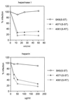
Dose-dependent reduction of infection of SK6 cells with reisolated S-ST (blood from pig 4071) and S-RT (blood from pig 4061) viruses and with S-ST virus produced in SK6 cells [SK6(S-ST)] after heparinase I treatment of cells or by addition of 200 μg of heparin per ml to the infection medium. Tissue culture wells (2 cm2) were infected with approximately 1,000 PFU of 4071 (S-ST) or 4061 (S-RT) virus and with 250 PFU of SK6(S-ST) virus. Blood samples taken on day 14 p.i. were tested. The number of plaques in wells was measured in a plaque assay, and the percent infection compared to that in control wells was calculated as described in Materials and Methods. Symbols represent the mean of two independent observations.
FIG. 3
Reduction of infection of primary swine kidney cells and lung macrophages with reisolated S-ST (blood from pig 4071) and S-RT (blood from pig 4061) viruses and with S-ST virus produced in SK6 cells [SK6(S-ST)] after treatment of cells with 20 mIU of heparinase I per ml or by addition of 200 μg of heparin per ml to the infection medium. Tissue culture wells (2 cm2) containing primary swine kidney cells were infected with approximately 300 PFU of 4071 (S-ST), 1,000 PFU of 4061 (S-RT), or 250 PFU of SK6(S-ST) virus. Wells with macrophages were infected with approximately 400 PFU of 4071 (S-ST), 1,000 PFU of 4061 (S-RT), or 400 PFU of SK6(S-ST) virus. Blood samples taken on day 14 p.i. were tested. The number of plaques in wells was measured in a macrophage plaque assay or in standard plaque assay (for primary kidney cells), and the percentage infection compared to control wells was calculated as described in Materials and Methods Bars represent the mean of three independent observations. Virus titers (log10 PFU per milliliter) in the blood of pigs 4071 and 4061, and SK6(S-ST) virus were measured in a macrophage plaque assay and in a standard plaque assay using cultured (SK6) or primary swine kidney (kidney) cells (table).
FIG. 4
Inhibition or stimulation of infection by ConA. Viruses were preincubated with different concentrations of ConA for 30 min at 37°C. Preincubation mixtures were used to infect 2-cm2 tissue culture wells with SK6 cells for 30 min at 37°C. After removal of virus and washing of cells, the cells were incubated with 100 mM MMP for 30 min at 37°C. Subsequently, they were washed and supplied with overlay medium and stained after 24 h of growth at 37°C. Symbols represent the mean number of plaques calculated from two independent observations.
FIG. 5
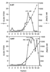
Heparin-Sepharose chromatography of Erns. Purified Erns S-ST or Erns S-RT was loaded onto the column at a concentration of 0 mM NaCl. Proteins were eluted by increasing the NaCl concentration stepwise. The fractions were assayed for Erns in an ELISA, and the NaCl concentration of these fractions was determined by measuring the osmolarity.
Similar articles
- Determinants of virulence of classical swine fever virus strain Brescia.
Van Gennip HG, Vlot AC, Hulst MM, De Smit AJ, Moormann RJ. Van Gennip HG, et al. J Virol. 2004 Aug;78(16):8812-23. doi: 10.1128/JVI.78.16.8812-8823.2004. J Virol. 2004. PMID: 15280489 Free PMC article. - Dimerization of glycoprotein E(rns) of classical swine fever virus is not essential for viral replication and infection.
van Gennip HG, Hesselink AT, Moormann RJ, Hulst MM. van Gennip HG, et al. Arch Virol. 2005 Nov;150(11):2271-86. doi: 10.1007/s00705-005-0569-y. Epub 2005 Jun 28. Arch Virol. 2005. PMID: 15986175 - Approaches to define the viral genetic basis of classical swine fever virus virulence.
Leifer I, Ruggli N, Blome S. Leifer I, et al. Virology. 2013 Apr 10;438(2):51-5. doi: 10.1016/j.virol.2013.01.013. Epub 2013 Feb 13. Virology. 2013. PMID: 23415391 Review. - Genetic variability and distribution of Classical swine fever virus.
Beer M, Goller KV, Staubach C, Blome S. Beer M, et al. Anim Health Res Rev. 2015 Jun;16(1):33-9. doi: 10.1017/S1466252315000109. Anim Health Res Rev. 2015. PMID: 26050570 Review.
Cited by
- Modulation of ADAM17 Levels by Pestiviruses Is Species-Specific.
Chen HW, Zaruba M, Dawood A, Düsterhöft S, Lamp B, Ruemenapf T, Riedel C. Chen HW, et al. Viruses. 2024 Oct 2;16(10):1564. doi: 10.3390/v16101564. Viruses. 2024. PMID: 39459898 Free PMC article. - Integrin αVβ3 mediates porcine deltacoronavirus infection and inflammatory response through activation of the FAK-PI3K-AKT-nf-κB signalling pathway.
Jin X, Wu X, Li Z, Hu Y, Xia L, Zu S, Zhang G, Hu H. Jin X, et al. Virulence. 2024 Dec;15(1):2407847. doi: 10.1080/21505594.2024.2407847. Epub 2024 Oct 5. Virulence. 2024. PMID: 39368071 Free PMC article. - Host cell factors involved in classical swine fever virus entry.
Lamothe-Reyes Y, Figueroa M, Sánchez O. Lamothe-Reyes Y, et al. Vet Res. 2023 Dec 1;54(1):115. doi: 10.1186/s13567-023-01238-x. Vet Res. 2023. PMID: 38041163 Free PMC article. Review. - Attachment, Entry, and Intracellular Trafficking of Classical Swine Fever Virus.
Guo X, Zhang M, Liu X, Zhang Y, Wang C, Guo Y. Guo X, et al. Viruses. 2023 Sep 3;15(9):1870. doi: 10.3390/v15091870. Viruses. 2023. PMID: 37766277 Free PMC article. Review. - Host Cell Receptors Implicated in the Cellular Tropism of BVDV.
Qi S, Wo L, Sun C, Zhang J, Pang Q, Yin X. Qi S, et al. Viruses. 2022 Oct 20;14(10):2302. doi: 10.3390/v14102302. Viruses. 2022. PMID: 36298858 Free PMC article. Review.
References
- Anonymous. Tissue culture methods and applications. New York, N.Y: Academic Press, Inc.; 1973.
- Becher P, Shannon A D, Tautz N, Thiel H-J. Molecular characterization of border disease virus, a pestivirus from sheep. Virology. 1994;198:542–551. - PubMed
Publication types
MeSH terms
Substances
LinkOut - more resources
Full Text Sources


