Murine cytomegalovirus CC chemokine homolog MCK-2 (m131-129) is a determinant of dissemination that increases inflammation at initial sites of infection - PubMed (original) (raw)
Comparative Study
Murine cytomegalovirus CC chemokine homolog MCK-2 (m131-129) is a determinant of dissemination that increases inflammation at initial sites of infection
N Saederup et al. J Virol. 2001 Oct.
Abstract
The murine cytomegalovirus CC chemokine homolog MCK-2 (m131-129) is an important determinant of dissemination during primary infection. Reduced peak levels of viremia at day 5 were followed by reduced levels of virus in salivary glands starting at day 7 when mck insertion (RM461) and point (RM4511) mutants were compared to mck-expressing viruses. A dramatic MCK-2-enhanced inflammation occurred at the inoculation site over the first few days of infection, preceding viremia. The data further reinforce the role of MCK-2 as a proinflammatory signal that recruits leukocytes to increase the efficiency of viral dissemination in the host.
Figures
FIG. 1
Schematic representation of mutant virus genomes. The top line represents a restriction map of the _Hin_dIII K, L, and J DNA fragments in murine CMV strain K181+, corresponding to nts 173170 to 195847 of the Smith strain genome (46) (GenBank accession number U68299). Restriction sites for _Hin_dIII and selected _Bss_HI, _Hpa_I, and _Bbr_PI sites are indicated above the line, and a 1-kbp scale marker is indicated by a double-headed arrow below the line. Open boxes with arrowheads depict the positions of viral ORFs, with m131 and m129 contributing the coding sequences for MCK-2 (35). m130 overlaps mck on the opposite DNA strand (46). Solid arrows depict transcripts (_ie_1, _ie_3, _ie_2, mck, and _sgg_1) encoded by wild-type viruses. The mutations introduced into mutant viruses RM427+, RM4503, RM461, and RM4511 are depicted below the transcripts. The 3.9-kbp lacZ insert carried by RM427+ and RM461 (open box) is controlled by a 199-bp human CMV _ie_1-_ie_2 promoter fragment (shaded box) encompassing positions −219 to −19 relative to the transcription start site (8, 37, 55). The 1.7-kbp EGFP-puro insert in RM4503 and RM4511 (open box) is controlled by a 248-bp human CMV _ie_1-_ie_2 promoter fragment (hatched box) encompassing positions −242 to +7 relative to the transcription start site (59). The expanded region shows aa 25 to 30 of m131 and aa 142 to 147 of m130, including the two nucleotide point mutations (denoted by asterisks) introduced into RM4511, generating a new _Bbr_PI site (underlined) and altering the MCK-2 amino acid sequence (C27R and C28G; bold type). Wild-type strain K181+ nucleotide and amino acid sequences are shown at the bottom.
FIG. 2
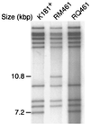
Restriction digestion analysis of RQ461 DNA. Autoradiograph of 32P-end-labeled _Hin_dIII fragments from K181+, RM461, and RQ461 DNA following electrophoretic separation on a 0.7% agarose gel.
FIG. 3
Restriction digestion analysis of RM4511 DNA. (A) Detection of the RM4511 mck mutation by _Bbr_PI digestion. Autoradiograph of _Bbr_PI-digested (Roche) virion DNA from parental RM427+, RM4503, and two isolates of RM4511 (RM4511.1 and RM4511.2) following electrophoretic separation on a 1% agarose gel and hybridization with a _Hin_dIII/_Afl_II fragment mck probe. (B) Detection of the EGFP-puro insert in RM4503 and RM4511. Autoradiograph of electrophoretically separated, _Bss_HII-digested 32P-end-labeled DNA fragments. (C) Autoradiograph of electrophoretically separated 32P-end-labeled _Hin_dIII fragments of DNA from RM427+, RM4503, and two isolates of RM4511.
FIG. 4
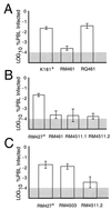
Evaluation of peak levels of viremia at 5 days after i.p. inoculation of BALB/c mice with 106 PFU. (A) Comparison of K181+, RM461, and RQ461. (B) Comparison of RM427+, RM461, RM4511.1, and RM4511.2. (C) Comparison of RM427+, RM4503, and RM4511.2. PBLs (106) were harvested and subjected to an infectious-center assay on NIH 3T3 cells. Bar height indicates the geometric mean, and vertical bars indicate standard deviations of the geometric mean. The shaded area indicates the limit of detection of virus.
FIG. 5
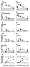
Replication patterns for RM461, RQ461, and K181+ in BALB/c mice. Experiment 1 (A, C, E, G, I, and K) shows virus titers in organs at 3, 5, 7, and 14 days postinoculation (four animals per group). Experiment 2 (B, D, F, H, J, and L) shows titers at 1, 2, 3, 5, 7, and 14 days postinoculation (five animals per group). Footpads of 3-week-old BALB/c mice were inoculated with 106 PFU. Titers in organ sonicates were determined by plaque assays on NIH 3T3 cells. Each symbol represents an individual mouse. K181+ (□), RM461 (▵), and RQ461 (▴) were the viruses used. The lines connect the geometric means for each virus (solid, K181+; dotted, RQ461; dashed, RM461). The shaded area indicates the limit of detection of virus.
FIG. 6
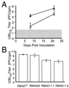
Dissemination of mck mutant viruses to salivary glands. (A) Levels of RM461 (▵) or RQ461 (▴) in the salivary glands at 8 and 21 days postinoculation of 100 PFU into footpads of 3-week-old BALB/c mice (four animals per group). (B) Levels of RM427+, RM4503, RM4511.1, and RM4511.2 in the salivary glands at 14 days postinoculation of 106 PFU into footpads of 3-week-old BALB/c mice (four animals per group). Titers of organ sonicates were determined by plaque assays on NIH 3T3 cells, and mean values were plotted. Vertical bars indicate standard deviations of the geometric mean. The shaded area indicates the limit of detection of virus.
FIG. 7
Time course analysis of footpad swelling. (A and B) Measurement of RM461 (▵)- or RQ461 (▴)-induced swelling following footpad inoculation of 3-week-old BALB/c mice in groups of five (A) or four (B) animals. (C) Measurement of RM4511.1 (▾)-, RM4511.2 (▿)-, RM4503 (•)-, or RM427+ (▪)-induced swelling following footpad inoculation of three 5-week-old mice. Mice were inoculated with 106 PFU of virus. Foot thickness was measured with a digital caliper at the times indicated after inoculation, and mean values were plotted. Vertical bars indicate standard deviations of the mean.
FIG. 8
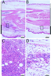
Inflammatory responses induced by RM461 and RQ461 following footpad inoculation. Tissues were harvested from an inoculated mouse foot 48 h after inoculation with RQ461 or RM461 (106 PFU in 3 μl of growth medium), formalin fixed, and decalcified; after embedding in paraffin, 5-μm sections were cut and stained with hematoxylin and eosin. (A) RQ461. (B) RM461. (C) RQ461. (D) RM461. The dorsal, internal, and ventral areas are denoted by white, grey, and black bars, respectively, on the sides of panels A and B. Areas boxed in panels A and B are magnified in panels C and D, respectively.
FIG. 9
Histopathological evaluation of edema and cellularity following footpad inoculation. Groups of five mice were inoculated in a single footpad with either RM461 or RQ461 as described in the legend to Fig. 8, and foot sections were collected for analysis at days 2, 3, and 7 postinoculation. Sections from each foot were evaluated for inflammatory changes at low-power magnification by light microscopy (×40) and assigned numerical values (see the text) for levels of cellularity (A) and edema (B). Bars correspond to the mean values for the dorsal (open), internal (grey), and ventral (black) areas and are depicted to appreciate the score for an area as well as a total score (maximum of 15) that incorporates the evaluation of all three areas.
Similar articles
- The r131 gene of rat cytomegalovirus encodes a proinflammatory CC chemokine homolog which is essential for the production of infectious virus in the salivary glands.
Kaptein SJ, van Cleef KW, Gruijthuijsen YK, Beuken EV, van Buggenhout L, Beisser PS, Stassen FR, Bruggeman CA, Vink C. Kaptein SJ, et al. Virus Genes. 2004 Aug;29(1):43-61. doi: 10.1023/B:VIRU.0000032788.53592.7c. Virus Genes. 2004. PMID: 15215683 - The murine cytomegalovirus chemokine homolog, m131/129, is a determinant of viral pathogenicity.
Fleming P, Davis-Poynter N, Degli-Esposti M, Densley E, Papadimitriou J, Shellam G, Farrell H. Fleming P, et al. J Virol. 1999 Aug;73(8):6800-9. doi: 10.1128/JVI.73.8.6800-6809.1999. J Virol. 1999. PMID: 10400778 Free PMC article. - Fatal attraction: cytomegalovirus-encoded chemokine homologs.
Saederup N, Mocarski ES Jr. Saederup N, et al. Curr Top Microbiol Immunol. 2002;269:235-56. doi: 10.1007/978-3-642-59421-2_14. Curr Top Microbiol Immunol. 2002. PMID: 12224512 - Murine Cytomegalovirus MCK-2 Facilitates In Vivo Infection Transfer from Dendritic Cells to Salivary Gland Acinar Cells.
Ma J, Bruce K, Stevenson PG, Farrell HE. Ma J, et al. J Virol. 2021 Aug 10;95(17):e0069321. doi: 10.1128/JVI.00693-21. Epub 2021 Aug 10. J Virol. 2021. PMID: 34132572 Free PMC article. - The viral chemokine MCK-2 of murine cytomegalovirus promotes infection as part of a gH/gL/MCK-2 complex.
Wagner FM, Brizic I, Prager A, Trsan T, Arapovic M, Lemmermann NA, Podlech J, Reddehase MJ, Lemnitzer F, Bosse JB, Gimpfl M, Marcinowski L, MacDonald M, Adler H, Koszinowski UH, Adler B. Wagner FM, et al. PLoS Pathog. 2013;9(7):e1003493. doi: 10.1371/journal.ppat.1003493. Epub 2013 Jul 25. PLoS Pathog. 2013. PMID: 23935483 Free PMC article.
Cited by
- Novel heparan sulfate-binding peptides for blocking herpesvirus entry.
Dogra P, Martin EB, Williams A, Richardson RL, Foster JS, Hackenback N, Kennel SJ, Sparer TE, Wall JS. Dogra P, et al. PLoS One. 2015 May 18;10(5):e0126239. doi: 10.1371/journal.pone.0126239. eCollection 2015. PLoS One. 2015. PMID: 25992785 Free PMC article. - Epstein-Barr virus-encoded BILF1 is a constitutively active G protein-coupled receptor.
Paulsen SJ, Rosenkilde MM, Eugen-Olsen J, Kledal TN. Paulsen SJ, et al. J Virol. 2005 Jan;79(1):536-46. doi: 10.1128/JVI.79.1.536-546.2005. J Virol. 2005. PMID: 15596846 Free PMC article. - Multiple Autonomous Cell Death Suppression Strategies Ensure Cytomegalovirus Fitness.
Mandal P, Nagrani LN, Hernandez L, McCormick AL, Dillon CP, Koehler HS, Roback L, Alnemri ES, Green DR, Mocarski ES. Mandal P, et al. Viruses. 2021 Aug 27;13(9):1707. doi: 10.3390/v13091707. Viruses. 2021. PMID: 34578288 Free PMC article. - Murine Cytomegalovirus Infection Induces Susceptibility to EAE in Resistant BALB/c Mice.
Milovanovic J, Popovic B, Milovanovic M, Kvestak D, Arsenijevic A, Stojanovic B, Tanaskovic I, Krmpotic A, Arsenijevic N, Jonjic S, Lukic ML. Milovanovic J, et al. Front Immunol. 2017 Feb 27;8:192. doi: 10.3389/fimmu.2017.00192. eCollection 2017. Front Immunol. 2017. PMID: 28289417 Free PMC article. - The r131 gene of rat cytomegalovirus encodes a proinflammatory CC chemokine homolog which is essential for the production of infectious virus in the salivary glands.
Kaptein SJ, van Cleef KW, Gruijthuijsen YK, Beuken EV, van Buggenhout L, Beisser PS, Stassen FR, Bruggeman CA, Vink C. Kaptein SJ, et al. Virus Genes. 2004 Aug;29(1):43-61. doi: 10.1023/B:VIRU.0000032788.53592.7c. Virus Genes. 2004. PMID: 15215683
References
- Alam R, Kumar D, Anderson-Walters D, Forsythe P A. Macrophage inflammatory protein-1 alpha and monocyte chemoattractant peptide-1 elicit immediate and late cutaneous reactions and activate murine mast cells in vivo. J Immunol. 1994;152:1298–1303. - PubMed
- Baggiolini M, Dewald B, Moser B. Human chemokines: an update. Annu Rev Immunol. 1997;15:675–705. - PubMed
- Baggiolini M, Loetscher P. Chemokines in inflammation and immunity. Immunol Today. 2000;21:418–420. - PubMed
Publication types
MeSH terms
Substances
Grants and funding
- K08 AI 01638/AI/NIAID NIH HHS/United States
- R01 AI030363/AI/NIAID NIH HHS/United States
- AI33852/AI/NIAID NIH HHS/United States
- R21 AI030363/AI/NIAID NIH HHS/United States
- AI30363/AI/NIAID NIH HHS/United States
- R56 AI030363/AI/NIAID NIH HHS/United States
- T32 GM07328/GM/NIGMS NIH HHS/United States
- R01 AI033852/AI/NIAID NIH HHS/United States
LinkOut - more resources
Full Text Sources
Other Literature Sources

