Ectopic expression of retinoic acid early inducible-1 gene (RAE-1) permits natural killer cell-mediated rejection of a MHC class I-bearing tumor in vivo - PubMed (original) (raw)
Ectopic expression of retinoic acid early inducible-1 gene (RAE-1) permits natural killer cell-mediated rejection of a MHC class I-bearing tumor in vivo
A Cerwenka et al. Proc Natl Acad Sci U S A. 2001.
Abstract
In 1986, Kärre and colleagues reported that natural killer (NK) cells rejected an MHC class I-deficient tumor cell line (RMA-S) but they did not reject the same cell line if it expressed MHC class I (RMA). Based on this observation, they proposed the concept that NK cells provide immune surveillance for "missing self," e.g., they eliminate cells that have lost class I MHC antigens. This seminal observation predicted the existence of inhibitory NK cell receptors for MHC class I. Here, we present evidence that NK cells are able to reject tumors expressing MHC class I if the tumor expresses a ligand for NKG2D. Mock-transfected RMA cells resulted in tumor formation. In contrast, when RMA cells were transfected with the retinoic acid early inducible gene-1 gamma or delta (RAE-1), ligands for the activating receptor NKG2D, the tumors were rejected. The tumor rejection was mediated by NK cells, and not by CD1-restricted NK1.1(+) T cells. No T cell-mediated immunological memory against the parental tumor was generated in the animals that had rejected the RAE-1 transfected tumors, which succumbed to rechallenge with the parental RMA tumor. Therefore, NK cells are able to reject a tumor expressing RAE-1 molecules, despite expression of self MHC class I on the tumor, demonstrating the potential for NK cells to participate in immunity against class I-bearing malignancies.
Figures
Figure 1
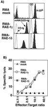
Ectopic expression of RAE-1 renders the MHC class I-expressing lymphoma cell line RMA susceptible to NK cell attack. (A) Mock-transfected RMA cells (Top) or RMA cells transfected with RAE-1γ or RAE-1δ (Middle and_Bottom_) were stained with the mNKG2D-Ig-FP (filled histograms, Left) or a control Ig fusion protein (CO-Ig-FP; open histograms, Left) and a PE-conjugated goat anti-human IgG second-step antibody. Transfectants were also stained with an H-2Db mAb (filled histograms,Right) or an isotype-matched control Ig (open histograms, Right). Cells were analyzed by flow cytometry. Data are displayed as histograms (x axis, fluorescence, 4-decade log scale; y axis, relative number of cells). (B) IL-2 activated NK cells from B6 mice were used as effectors in 4-h 51Cr-release assays at the indicated effector-to-target ratios. Targets were mock-transfected RMA cells (▴) or RMA cells transfected with RAE-1γ (□) or RAE-1δ (■). Results from a representative experiment of three independent experiments are shown.
Figure 2
RAE-1 expression on the RMA lymphoma causes tumor rejection in vivo. (A) B6 mice were injected i.p. with 1 × 10⁴ mock-transfected RMA cells (■) or RAE-1γ transfected RMA cells (□). Data were compiled from three independently performed experiments using six mice in each group. Significance was determined by the log rank test to compare survival curves; P ≤ 0.001,n = 18. (B) B6 mice were injected i.p. with 1 × 10⁴ mock-transfected RMA cells (■) or RAE-1δ transfected RMA cells (□). Survival data for six animals per group are shown. (C) B6 mice were injected i. p. with 1 × 10⁵ (Top), 1 × 10⁴ (Middle), or 1 × 10³ (Bottom) mock-transfected RMA cells (■) or RAE-1γ transfected RMA cells (□). Survival data for five animals per group (Top) or six animals per group (Middle and Bottom) are shown.
Figure 3
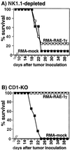
NK cells cause the rejection of RAE-1γ-transfected RMA tumors. (A) About 1 × 10⁴ mock-transfected RMA cells (■) or RMA cells transfected with RAE-1γ (□) were injected i.p. into B6 animals. On days −4 and −2 before tumor inoculation and weekly thereafter, recipient mice were treated with anti-NK1.1 mAb i.p. (200 μg per mouse). Data were compiled from two independent experiments using six mice in each group. Survival curves were not significantly different as determined by the log rank test to compare survival curves, P ≤ 0.442, n = 12. (B) CD1-deficient mice on a C57BL/6 background were injected i.p. with 1 × 10⁴ mock-transfected RMA cells (■) or RMA cells transfected with RAE-1γ (□). Survival data for six animals per group are shown.
Figure 4
Simultaneous inoculation of RMA-mock and RMA-RAE-1γ results in tumor formation. Mock-transfected RMA cells (1 × 10⁴, ⋄), RMA cells transfected with RAE-1γ (1 × 10⁴, ■) or a 1:1 mixture of mock-transfected RMA cells and RMA cells transfected with RAE-1γ (●) were injected i.p. into B6 animals. Survival data for six animals per group are shown.
Figure 5
No tumor-specific T cell immunological memory is observed in mice that have rejected RAE-1γ transfected tumors. B6 animals that had rejected 1 × 10⁴ RMA cells transfected with RAE-1γ (⋄) were injected with 1 × 10⁴ mock-transfected RMA cells at 3 months after the primary tumor inoculation. In parallel, naïve, unprimed animals were injected with 1 × 10⁴ mock-transfected RMA cells (■). Representative survival data for 12 animals per group are shown.
Similar articles
- Cutting edge: tumor rejection mediated by NKG2D receptor-ligand interaction is dependent upon perforin.
Hayakawa Y, Kelly JM, Westwood JA, Darcy PK, Diefenbach A, Raulet D, Smyth MJ. Hayakawa Y, et al. J Immunol. 2002 Nov 15;169(10):5377-81. doi: 10.4049/jimmunol.169.10.5377. J Immunol. 2002. PMID: 12421908 - Blastocyst MHC, a putative murine homologue of HLA-G, protects TAP-deficient tumor cells from natural killer cell-mediated rejection in vivo.
Tajima A, Tanaka T, Ebata T, Takeda K, Kawasaki A, Kelly JM, Darcy PK, Vance RE, Raulet DH, Kinoshita K, Okumura K, Smyth MJ, Yagita H. Tajima A, et al. J Immunol. 2003 Aug 15;171(4):1715-21. doi: 10.4049/jimmunol.171.4.1715. J Immunol. 2003. PMID: 12902470 - T cells gene-engineered with DAP12 mediate effector function in an NKG2D-dependent and major histocompatibility complex-independent manner.
Teng MW, Kershaw MH, Hayakawa Y, Cerutti L, Jane SM, Darcy PK, Smyth MJ. Teng MW, et al. J Biol Chem. 2005 Nov 18;280(46):38235-41. doi: 10.1074/jbc.M505331200. Epub 2005 Sep 16. J Biol Chem. 2005. PMID: 16169855 - The innate immune response to tumors and its role in the induction of T-cell immunity.
Diefenbach A, Raulet DH. Diefenbach A, et al. Immunol Rev. 2002 Oct;188:9-21. doi: 10.1034/j.1600-065x.2002.18802.x. Immunol Rev. 2002. PMID: 12445277 Review. - The biology of NK cells and their receptors affects clinical outcomes after hematopoietic cell transplantation (HCT).
Foley B, Felices M, Cichocki F, Cooley S, Verneris MR, Miller JS. Foley B, et al. Immunol Rev. 2014 Mar;258(1):45-63. doi: 10.1111/imr.12157. Immunol Rev. 2014. PMID: 24517425 Free PMC article. Review.
Cited by
- Fratricide of natural killer cells dressed with tumor-derived NKG2D ligand.
Nakamura K, Nakayama M, Kawano M, Amagai R, Ishii T, Harigae H, Ogasawara K. Nakamura K, et al. Proc Natl Acad Sci U S A. 2013 Jun 4;110(23):9421-6. doi: 10.1073/pnas.1300140110. Epub 2013 May 20. Proc Natl Acad Sci U S A. 2013. PMID: 23690625 Free PMC article. - Impairment of NKG2D-Mediated Tumor Immunity by TGF-β.
Lazarova M, Steinle A. Lazarova M, et al. Front Immunol. 2019 Nov 15;10:2689. doi: 10.3389/fimmu.2019.02689. eCollection 2019. Front Immunol. 2019. PMID: 31803194 Free PMC article. Review. - CLEC5a-directed bispecific antibody for effective cellular phagocytosis.
Kedage V, Ellerman D, Fei M, Liang WC, Zhang G, Cheng E, Zhang J, Chen Y, Huang H, Lee WP, Wu Y, Yan M. Kedage V, et al. MAbs. 2022 Jan-Dec;14(1):2040083. doi: 10.1080/19420862.2022.2040083. MAbs. 2022. PMID: 35293277 Free PMC article. - NKG2D recognition and perforin effector function mediate effective cytokine immunotherapy of cancer.
Smyth MJ, Swann J, Kelly JM, Cretney E, Yokoyama WM, Diefenbach A, Sayers TJ, Hayakawa Y. Smyth MJ, et al. J Exp Med. 2004 Nov 15;200(10):1325-35. doi: 10.1084/jem.20041522. J Exp Med. 2004. PMID: 15545356 Free PMC article. - Suppression of tumor formation in lymph nodes by L-selectin-mediated natural killer cell recruitment.
Chen S, Kawashima H, Lowe JB, Lanier LL, Fukuda M. Chen S, et al. J Exp Med. 2005 Dec 19;202(12):1679-89. doi: 10.1084/jem.20051473. Epub 2005 Dec 13. J Exp Med. 2005. PMID: 16352740 Free PMC article.
References
- Biron C A, Nguyen K B, Pien G C, Cousens L P, Salazar-Mather T P. Annu Rev Immunol. 1999;17:189–220. - PubMed
- Lanier L L. Curr Opin Immunol. 1995;7:626–631. - PubMed
- Yu Y Y L, Kumar V, Bennett M. Annu Rev Immunol. 1992;10:189–214. - PubMed
- Kärre K, Ljunggren H G, Piontek G, Kiessling R. Nature (London) 1986;319:675–678. - PubMed
Publication types
MeSH terms
Substances
LinkOut - more resources
Full Text Sources
Other Literature Sources
Research Materials
Miscellaneous


