The Nsp1p carboxy-terminal domain is organized into functionally distinct coiled-coil regions required for assembly of nucleoporin subcomplexes and nucleocytoplasmic transport - PubMed (original) (raw)
The Nsp1p carboxy-terminal domain is organized into functionally distinct coiled-coil regions required for assembly of nucleoporin subcomplexes and nucleocytoplasmic transport
S M Bailer et al. Mol Cell Biol. 2001 Dec.
Abstract
Nucleoporin Nsp1p, which has four predicted coiled-coil regions (coils 1 to 4) in the essential carboxy-terminal domain, is unique in that it is part of two distinct nuclear pore complex (NPC) subcomplexes, Nsp1p-Nup57p-Nup49p-Nic96p and Nsp1p-Nup82p-Nup159p. As shown by in vitro reconstitution, coiled-coil region 2 (residues 673 to 738) is sufficient to form heterotrimeric core complexes and can bind either Nup57p or Nup82p. Accordingly, interaction of Nup82p with Nsp1p coil 2 is competed by excess Nup57p. Strikingly, coil 3 and 4 mutants are still assembled into the core Nsp1p-Nup57p-Nup49p complex but no longer associate with Nic96p. Consistently, the Nsp1p-Nup57p-Nup49p core complex dissociates from the nuclear pores in nsp1 coil 3 and 4 mutant cells, and as a consequence, defects in nuclear protein import are observed. Finally, the nsp1-L640S temperature-sensitive mutation, which maps in coil 1, leads to a strong nuclear mRNA export defect. Thus, distinct coiled-coil regions within Nsp1p-C have separate functions that are related to the assembly of different NPC subcomplexes, nucleocytoplasmic transport, and incorporation into the nuclear pores.
Figures
FIG. 1
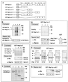
In vitro reconstitution of two different heterotrimeric Nsp1p-containing complexes. (A) Schematic drawing of GST–Nsp1p-C constructs GST-Nsp1p coil 1 (positions 591 to 672), GST-Nsp1p coil 2 (673 to 738), GST-Nsp1p coils 2 and 3 (673 to 781), GST-Nsp1p coils 1 and 2 (591 to 738), and GST-Nsp1p coils 3 and 4 (728 to 823). (B) Purified GST–Nsp1p-C (lane 1) was mixed with urea-treated bacterial lysates containing His6-Nup57p (lane 2). Proteins affinity purified via GSH-Sepharose (lane 4) were separated from unbound proteins (lane 3). (C) Purified GST–Nsp1p-C was mixed with urea-treated bacterial lysates containing His6-Nup49p (lane 1), His6-Nup57p (lane 2), or a mixture of both (lane 3). (D) Purified GST–Nsp1p-C (lanes 1 to 3) or GST alone (lane 4) was mixed with urea-treated bacterial lysate containing His6-Nup82p (lane 1), His6–Nup159p-C (lane 2 and 4), or a mixture of both (lane 3). (E) GST-tagged fragments of Nsp1p-C (as shown in panel A) were mixed with urea-treated bacterial lysate containing His6-Nup57p or His6-Nup82p. Open circles represent GST fusion proteins, filled triangles represent Nup57p, and open triangles represent Nup82p. (F) GST–Nsp1p-C (lane 1), GST-Nsp1p coils 2 and 3 (lane 2), GST-Nsp1p coil 2 (lane 3), or GST alone (lane 4) was mixed with urea-treated bacterial lysate containing His6-Nup49p and His6-Nup57p. (G) GST-Nsp1p coil 2 (lanes 1 to 3) was mixed with urea-treated bacterial lysate containing His6-Nup82p (lane 1), His6–Nup159p-C (lane 2), or a mixture of both (lane 3). For panels B to G, dialysis of the protein mixture was followed by affinity purification of the reconstituted GST-Nsp1p subcomplexes or of GST on glutathione-Sepharose. Proteins eluted from the column were analyzed by SDS-PAGE and Coomassie staining or Western blotting with anti-His, anti-Nsp1p, or anti-GST antibodies.
FIG. 2
Nup82p and Nup57p compete for binding to Nsp1p-C. (A) Schematic drawing of the competition assay. (B) GST-Nup57p (lanes 1 and 2, indicated by open circles) or GST–Nsp1p-C (lane 3, indicated by an open circle) was mixed with urea-treated bacterial lysate containing His6-Nup82p and His6–Nsp1p-C (lane 1) or only His6-Nup82p (lane 2 and 3). In vitro reconstitution was performed as described in the legend to Fig. 1. Proteins eluted from the glutathione-Sepharose or unbound proteins were analyzed by SDS-PAGE, followed by silver staining or Western blotting with anti-Nup57p, anti-Nup82p, or anti-Nsp1p antibodies. GST-Nsp1p, used in lane 3, is not shown in the Western blot. The band seen in the anti-Nup57p Western blot (lane 3) results from the first incubation of this membrane with anti-Nup82p antibodies and thus represents His6-Nup82p. (C) GST–Nsp1p-C was mixed with bacterial lysates containing His6-Nup82p (lane 1), both His6-Nup82p lysate and increasing amounts of purified His6-Nup57p (lanes 2 to 6), or His6-Nup57p alone (lane 7). In vitro reconstitution was performed as described in the legend to Fig. 1, and proteins eluted from glutathione-Sepharose were analyzed by SDS-PAGE and Coomassie staining or Western blotting with anti-His antibodies.
FIG. 3
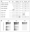
Mutational analysis of the various coiled-coil regions within Nsp1p-C. (A) Schematic drawings of Nsp1p-C (591–823) (map 1), Nsp1p-C (630–823) (map 2), nsp1-(638–823) (map 3), nsp1-(642–823) (map 4), nsp1-L640S (map 5), nsp1 ts_Δ_4 (map 6), nsp1 ts18 (map 7), and nsp1-ala6 (map 8). (B) Growth properties of Nsp1p-C and nsp1 ts cells as indicated in panel A. In each case, the _nsp1_Δ strain was complemented by a plasmid expressing the corresponding mutant (see Table 2). Precultures were diluted in 10−1 steps, and equivalent amounts of cells were dropped on YPD plates and incubated at 23, 30, or 37°C for 3 to 4 days.
FIG. 4
Roles of the various coiled-coil regions of Nsp1p-C in nucleocytoplasmic transport. (A) Nuclear poly(A)+ RNA export. NSP1-C or nsp1-L640S, nsp1-ala6, and nsp1 ts18 cells were grown at 23°C before a shift to 37°C for 0 min, 30 min, or 3 h. The localization of poly(A)+ RNA was analyzed by in situ hybridization with a Cy3-labeled oligo(dT) probe. DNA was visualized by 4′,6′-diamidino-2-phenylindole (DAPI) staining. (B) NPC localization of GFP-Nup82p or GFP-Nup159p in nsp1-L640S cells as revealed by fluorescence microscopy. Cells were grown in selective media at 23°C or shifted to 37°C for 2 h. (C) Nuclear protein import. NSP1-C, nsp1-L640S, nsp1-ala6, and nsp1 ts18 cells expressing GFP-Npl3p were grown in selective media at 23°C or shifted to 37°C for 3 h and analyzed by fluorescence microscopy. In each case, the _nsp1_Δ strain was complemented by a plasmid expressing the corresponding mutant (see Table 2).
FIG. 5
Nsp1p coils 3 and 4 are required for docking of the Nsp1p-Nup57p-Nup49p complex to the NPC. (A) Schematic drawing of nsp1 ts18. (B) Localization of GFP-NSP1-C and GFP-nsp1 ts18 expressed in _nsp1_Δ cells. Bars, 4 μm. (C) Localization of GFP-Nic96p, GFP-Nup57p, GFP-Nup82p, and GFP-Nup159p in _nsp1_Δ/_nupX_Δ strains (see Table 1) complemented by two plasmids expressing NSP1-C or nsp1 ts18 cells and GFP-NupXp. Cells were grown in selective media at 23°C and analyzed by fluorescence microscopy or Nomarski optics. Bars, 4 μm. (D) Affinity purification of ProtA–Nsp1p-C and ProtA-nsp1 ts18 expressed in _nsp1_Δ cells. Whole-cell lysates (lanes 1 and 4) or proteins eluted from IgG-Sepharose (lanes 2 and 3) were analyzed by SDS-PAGE and Coomassie staining or by Western blotting with anti-Nup159p, anti-Nic96p, anti-Nup82p, anti-Nup57p, and anti-ProtA antibodies. The positions of ProtA fusion proteins are indicated by open circles, and the positions of copurifying proteins are indicated by asterisks.
FIG. 6
Model of interaction of Nup82p-Nup159p and Nup57p-Nup49p-Nic96p, respectively, with Nsp1p-C.
Similar articles
- Functional interaction of Nic96p with a core nucleoporin complex consisting of Nsp1p, Nup49p and a novel protein Nup57p.
Grandi P, Schlaich N, Tekotte H, Hurt EC. Grandi P, et al. EMBO J. 1995 Jan 3;14(1):76-87. doi: 10.1002/j.1460-2075.1995.tb06977.x. EMBO J. 1995. PMID: 7828598 Free PMC article. - In vitro reconstitution of a heterotrimeric nucleoporin complex consisting of recombinant Nsp1p, Nup49p, and Nup57p.
Schlaich NL, Häner M, Lustig A, Aebi U, Hurt EC. Schlaich NL, et al. Mol Biol Cell. 1997 Jan;8(1):33-46. doi: 10.1091/mbc.8.1.33. Mol Biol Cell. 1997. PMID: 9017593 Free PMC article. - A novel nuclear pore protein Nup82p which specifically binds to a fraction of Nsp1p.
Grandi P, Emig S, Weise C, Hucho F, Pohl T, Hurt EC. Grandi P, et al. J Cell Biol. 1995 Sep;130(6):1263-73. doi: 10.1083/jcb.130.6.1263. J Cell Biol. 1995. PMID: 7559750 Free PMC article. - Pharmacological interference with protein-protein interactions mediated by coiled-coil motifs.
Strauss HM, Keller S. Strauss HM, et al. Handb Exp Pharmacol. 2008;(186):461-82. doi: 10.1007/978-3-540-72843-6_19. Handb Exp Pharmacol. 2008. PMID: 18491064 Review. - [Nuclear transport of hnRNP and mRNA].
Taura T, Siomi H, Siomi MC. Taura T, et al. Tanpakushitsu Kakusan Koso. 2000 Oct;45(14):2378-87. Tanpakushitsu Kakusan Koso. 2000. PMID: 11051839 Review. Japanese. No abstract available.
Cited by
- Scaffold nucleoporins Nup188 and Nup192 share structural and functional properties with nuclear transport receptors.
Andersen KR, Onischenko E, Tang JH, Kumar P, Chen JZ, Ulrich A, Liphardt JT, Weis K, Schwartz TU. Andersen KR, et al. Elife. 2013 Jun 11;2:e00745. doi: 10.7554/eLife.00745. Elife. 2013. PMID: 23795296 Free PMC article. - The yeast nuclear pore complex and transport through it.
Aitchison JD, Rout MP. Aitchison JD, et al. Genetics. 2012 Mar;190(3):855-83. doi: 10.1534/genetics.111.127803. Genetics. 2012. PMID: 22419078 Free PMC article. - Ring cycle for dilating and constricting the nuclear pore.
Solmaz SR, Blobel G, Melcák I. Solmaz SR, et al. Proc Natl Acad Sci U S A. 2013 Apr 9;110(15):5858-63. doi: 10.1073/pnas.1302655110. Epub 2013 Mar 11. Proc Natl Acad Sci U S A. 2013. PMID: 23479651 Free PMC article. - Disorder in the nuclear pore complex: the FG repeat regions of nucleoporins are natively unfolded.
Denning DP, Patel SS, Uversky V, Fink AL, Rexach M. Denning DP, et al. Proc Natl Acad Sci U S A. 2003 Mar 4;100(5):2450-5. doi: 10.1073/pnas.0437902100. Epub 2003 Feb 25. Proc Natl Acad Sci U S A. 2003. PMID: 12604785 Free PMC article. - The nuclear pore complex has entered the atomic age.
Brohawn SG, Partridge JR, Whittle JR, Schwartz TU. Brohawn SG, et al. Structure. 2009 Sep 9;17(9):1156-68. doi: 10.1016/j.str.2009.07.014. Structure. 2009. PMID: 19748337 Free PMC article. Review.
References
- Bailer S M, Balduf C, Katahira J, Podtelejnikov A, Rollenhagen C, Mann M, Pante N, Hurt E. Nup116p associates with the Nup82p-Nsp1p-Nup159p nucleoporin complex. J Biol Chem. 2000;275:23540–23548. - PubMed
Publication types
MeSH terms
Substances
LinkOut - more resources
Full Text Sources
Molecular Biology Databases
Research Materials


