Insulin-regulated release from the endosomal recycling compartment is regulated by budding of specialized vesicles - PubMed (original) (raw)
Insulin-regulated release from the endosomal recycling compartment is regulated by budding of specialized vesicles
M A Lampson et al. Mol Biol Cell. 2001 Nov.
Free PMC article
Abstract
In several cell types, specific membrane proteins are retained intracellularly and rapidly redistributed to the surface in response to stimulation. In fat and muscle, the GLUT4 glucose transporter is dynamically retained because it is rapidly internalized and slowly recycled to the plasma membrane. Insulin increases the recycling of GLUT4, resulting in a net translocation to the surface. We have shown that fibroblasts also have an insulin-regulated recycling mechanism. Here we show that GLUT4 is retained within the transferrin receptor-containing general endosomal recycling compartment in Chinese hamster ovary (CHO) cells rather than being segregated to a specialized, GLUT4-recycling compartment. With the use of total internal reflection microscopy, we demonstrate that the TR and GLUT4 are transported from the pericentriolar recycling compartment in separate vesicles. These data provide the first functional evidence for the formation of distinct classes of vesicles from the recycling compartment. We propose that GLUT4 is dynamically retained within the endosomal recycling compartment in CHO cells because it is concentrated in vesicles that form more slowly than those that transport TR. In 3T3-L1 adipocytes, cells that naturally express GLUT4, we find that GLUT4 is partially segregated to a separate compartment that is inaccessible to the TR. We present a model for the formation of this specialized compartment in fat cells, based on the general mechanism described in CHO cells, which may explain the increased retention of GLUT4 and its insulin-induced translocation in fat cells.
Figures
Figure 1
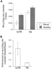
Recycling of vpTR and the TR is not affected by GLUT4 expression. (A) Cells expressing HA-GLUT4-GFP and either vpTR or the TR were loaded to steady state with 125I-Tf and allowed to efflux for times up to 10 min, with or without insulin. The recycling rate constant of the TR or vpTR was calculated as the slope of the natural log of the percentage of radioactivity cell associated versus time. Each bar represents the average of two experiments. Error bars show the difference between the two. (B) The ratio of the recycling rates (insulin/basal) is plotted for the TR and vpTR, for the experiments shown in A.
Figure 2
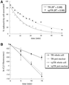
vpTR and the TR recycle as single kinetic pools from a perinuclear compartment. (A) Cells expressing either vpTR or the TR were loaded to steady state with 125I-Tf and allowed to efflux for times up to 50 min. The percentage of radioactivity remaining cell associated is plotted as a function of efflux time. Both curves are fit with single exponential decays. (B) Cells expressing HA-GLUT4-GFP and either vpTR or the TR were loaded to steady state with Cy3-Tf and allowed to efflux for times up to 15 min. The Cy3 fluorescence signal was summed either over the whole cell or in the perinuclear compartment only. At each time point, the Cy3 fluorescence intensity is normalized to the intensity at t = 0, and the natural log of this fraction is plotted versus time. Error bars are differences between duplicate dishes.
Figure 3
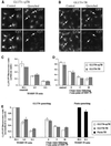
The GLUT4 retention compartment is accessible to both vpTR and the TR in CHO cells. (A and B) Cells expressing HA-GLUT4-GFP and either vpTR (A) or the TR (B) were incubated for 40 min with Cy3 anti-HA at 37°C, chased for 20 min with HRP-Tf, and processed for the DAB reaction. For control cells 1 mg/ml excess unconjugated Tf was included with the HRP-Tf to block specific binding of HRP-Tf. GFP and Cy3 fluorescence is shown. Arrows indicate the GLUT4 retention compartment in both control and quenched cells. Scale bar, 10 μm. (C) Cells were treated as in A and B and the Cy3/GFP ratio was calculated. Data are shown for a 50:1 ratio of unconjugated Tf/HRP-Tf (control), a 1:5 ratio of Tf/HRP-Tf, and for HRP-Tf only (maximal quenching). Each bar represents the mean of duplicate dishes, with eight fields averaged for each dish (>20 cells per field). Error bars represent the difference between the two dishes. (D) Cells were incubated for 40 min with Cy3 anti-HA, pulsed with HRP-Tf for 5 min, chased for the indicated times, and processed for the DAB reaction. The Cy3/GFP ratio was calculated and averaged over duplicate dishes as in C. The Cy3/GFP ratio is not comparable between the experiments in C and D. (E) Data were averaged over multiple experiments, performed as in C and D. To combine data from different experiments, the Cy3/GFP ratio was normalized to the control cells for each experiment. Each bar represents the mean (±SEM) of at least four experiments. Experiments with cells expressing the TR and furin were performed as in C.
Figure 4
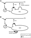
Models for retention in the ERC. (A) Two classes of vesicles bud from the ERC. The TR recycles in rapidly budding vesicles, and GLUT4 and vpTR are concentrated in separate, slowly budding vesicles. Insulin increases the budding of the slow vesicles. (B) Only one class of vesicles buds from the ERC. GLUT4 and vpTR are retained in the ERC by exclusion from these vesicles. The slow recycling of GLUT4 and vpTR is due to inefficient retention. Insulin releases the retention, allowing GLUT4 and vpTR to recycle with the TR. SE, sorting endosome; PM, plasma membrane.
Figure 5
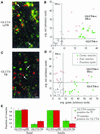
The TR is segregated from GLUT4 in transport vesicles imaged by TIR-FM. Cells expressing HA-GLUT4-GFP and either vpTR (A and B) or the TR (C and D) were loaded with Cy3-Tf and images were acquired by TIR-FM. (A and C) Images show structures containing GLUT4 (green), Tf (red), or both (yellow). Structures in A do not all contain the same ratio of vpTR to GLUT4; therefore, they do not all appear completely yellow. Scale bar, 1 μm. (B and D) Moving vesicles were selected for either GLUT4 content (solid green circles) or Tf content (open red circles). For each vesicle the average green intensity is plotted against the average red intensity, calculated over all the pixels in the vesicle. The intensity units are not comparable between the two plots. Spots randomly placed throughout the image represent background (×). Lines indicate threshold intensities, calculated from the random spots, to separate signal from background. Based on these thresholds, the quadrants are labeled as GLUT4(+) or Tf(+) to indicate where the intensities are higher than background. (E) The fraction of GLUT4 vesicles positive for Tf and Tf vesicles positive for GLUT4 is shown for the two cell types, both with and without insulin. The fractions are averaged over multiple videos in all cases, with at least 15 vesicles analyzed from each video. Error bars represent either the SEM of three or more determinations or the difference between two determinations.
Figure 6
Transport vesicles imaged by TIR-FM fuse with the plasma membrane. Cells expressing HA-GLUT4-GFP (A), the TR (B), or vpTR (C) were imaged by TIR-FM. Cells expressing the TR or vpTR were first loaded with Alexa488-Tf. Sequential frames, acquired at a rate of 6–10 frames/s, are shown for each probe. Arrows indicate vesicles that fuse in the sequence of frames shown. Asterisks mark frames showing the increase in area and decrease in peak intensity characteristic of fusion. Scale bar, 1 μm.
Figure 7
The GLUT4 retention compartment is only partially accessible to the TR in 3T3-L1 adipocytes. 3T3-L1 adipocytes expressing HA-GLUT4-GFP and either vpTR or the TR were incubated with Cy3 anti-HA, with or without HRP-Tf, and processed for the DAB reaction. The Cy3/GFP ratio (mean±SEM) was calculated and averaged over multiple cells. For both vpTR and the TR, the Cy3/GFP ratio with HRP-Tf is significantly different from the ratio with no HRP-Tf with p < 0.01 (heteroscedastic, two-tailed Student's t test).
Figure 8
Model for the retention pathway in adipocytes. VpTR, the TR, and GLUT4 are internalized from the plasma membrane and delivered to the ERC. Two classes of vesicles bud from the ERC. The TR recycles rapidly in one class of vesicle. In fibroblasts, vesicles containing vpTR and GLUT4 recycle slowly from the ERC, whereas in adipocytes, these vesicles are tethered inside the cell, forming a separate retention compartment. SE, sorting endosome; PM, plasma membrane.
Similar articles
- GLUT4 retention in adipocytes requires two intracellular insulin-regulated transport steps.
Zeigerer A, Lampson MA, Karylowski O, Sabatini DD, Adesnik M, Ren M, McGraw TE. Zeigerer A, et al. Mol Biol Cell. 2002 Jul;13(7):2421-35. doi: 10.1091/mbc.e02-02-0071. Mol Biol Cell. 2002. PMID: 12134080 Free PMC article. - Compartment ablation analysis of the insulin-responsive glucose transporter (GLUT4) in 3T3-L1 adipocytes.
Livingstone C, James DE, Rice JE, Hanpeter D, Gould GW. Livingstone C, et al. Biochem J. 1996 Apr 15;315 ( Pt 2)(Pt 2):487-95. doi: 10.1042/bj3150487. Biochem J. 1996. PMID: 8615819 Free PMC article. - Role of EHD1 and EHBP1 in perinuclear sorting and insulin-regulated GLUT4 recycling in 3T3-L1 adipocytes.
Guilherme A, Soriano NA, Furcinitti PS, Czech MP. Guilherme A, et al. J Biol Chem. 2004 Sep 17;279(38):40062-75. doi: 10.1074/jbc.M401918200. Epub 2004 Jul 9. J Biol Chem. 2004. PMID: 15247266 - Compartment-ablation studies of GLUT4 distribution in adipocytes: evidence for multiple intracellular pools.
Millar CA, Campbell LC, Cope DL, Melvin DR, Powell KA, Gould GW. Millar CA, et al. Biochem Soc Trans. 1997 Aug;25(3):974-7. doi: 10.1042/bst0250974. Biochem Soc Trans. 1997. PMID: 9388584 Review. - Moving GLUT4: the biogenesis and trafficking of GLUT4 storage vesicles.
Rea S, James DE. Rea S, et al. Diabetes. 1997 Nov;46(11):1667-77. doi: 10.2337/diab.46.11.1667. Diabetes. 1997. PMID: 9356011 Review.
Cited by
- Cortactin, an actin binding protein, regulates GLUT4 translocation via actin filament remodeling.
Nazari H, Khaleghian A, Takahashi A, Harada N, Webster NJ, Nakano M, Kishi K, Ebina Y, Nakaya Y. Nazari H, et al. Biochemistry (Mosc). 2011 Nov;76(11):1262-9. doi: 10.1134/S0006297911110083. Biochemistry (Mosc). 2011. PMID: 22117553 Free PMC article. - Αvβ3-integrin-mediated adhesion is regulated through an AAK1L- and EHD3-dependent rapid-recycling pathway.
Waxmonsky NC, Conner SD. Waxmonsky NC, et al. J Cell Sci. 2013 Aug 15;126(Pt 16):3593-601. doi: 10.1242/jcs.122465. Epub 2013 Jun 18. J Cell Sci. 2013. PMID: 23781025 Free PMC article. - Transcellular migration of leukocytes is mediated by the endothelial lateral border recycling compartment.
Mamdouh Z, Mikhailov A, Muller WA. Mamdouh Z, et al. J Exp Med. 2009 Nov 23;206(12):2795-808. doi: 10.1084/jem.20082745. Epub 2009 Nov 2. J Exp Med. 2009. PMID: 19887395 Free PMC article. - Resolving vesicle fusion from lysis to monitor calcium-triggered lysosomal exocytosis in astrocytes.
Jaiswal JK, Fix M, Takano T, Nedergaard M, Simon SM. Jaiswal JK, et al. Proc Natl Acad Sci U S A. 2007 Aug 28;104(35):14151-6. doi: 10.1073/pnas.0704935104. Epub 2007 Aug 21. Proc Natl Acad Sci U S A. 2007. PMID: 17715060 Free PMC article. - GLUT4 is retained by an intracellular cycle of vesicle formation and fusion with endosomes.
Karylowski O, Zeigerer A, Cohen A, McGraw TE. Karylowski O, et al. Mol Biol Cell. 2004 Feb;15(2):870-82. doi: 10.1091/mbc.e03-07-0517. Epub 2003 Oct 31. Mol Biol Cell. 2004. PMID: 14595108 Free PMC article.
References
- Axelrod D, Hellen EH, Fulbright RM. Total internal reflection fluorescence. In: Lakowicz JR, editor. Topics in Fluorescence Spectroscopy. 3: Biochemical Applications. New York: Plenum Press; 1992. pp. 289–343.
- Bradbury NA, Bridges RJ. Role of membrane trafficking in plasma membrane solute transport. Am J Physiol. 1994;267:C1–24. - PubMed
- Cannon C, van Adelsberg J, Kelly S, Al-Awqati Q. Carbon-dioxide-induced exocytotic insertion of H+ pumps in turtle-bladder luminal membrane: role of cell pH and calcium. Nature. 1985;314:443–446. - PubMed
Publication types
MeSH terms
Substances
Grants and funding
- DK-52852/DK/NIDDK NIH HHS/United States
- R01 DK057689/DK/NIDDK NIH HHS/United States
- R01 DK052852/DK/NIDDK NIH HHS/United States
- R56 DK052852/DK/NIDDK NIH HHS/United States
- DK-57689/DK/NIDDK NIH HHS/United States
LinkOut - more resources
Full Text Sources
Medical
