Toxoplasma gondii infection of neurons induces neuronal cytokine and chemokine production, but gamma interferon- and tumor necrosis factor-stimulated neurons fail to inhibit the invasion and growth of T. gondii - PubMed (original) (raw)
Toxoplasma gondii infection of neurons induces neuronal cytokine and chemokine production, but gamma interferon- and tumor necrosis factor-stimulated neurons fail to inhibit the invasion and growth of T. gondii
D Schlüter et al. Infect Immun. 2001 Dec.
Abstract
The intracellular parasite Toxoplasma gondii has the capacity to persist in the brain within neurons. In this study we demonstrated that T. gondii infected murine cerebellar neurons in vitro and replicated within these cells. Stimulation with gamma interferon (IFN-gamma) and/or tumor necrosis factor (TNF) did not enable neurons to inhibit parasite invasion and replication. Cultured neurons constitutively produced interleukin 1 (IL-1), IL-6, macrophage inflammatory protein 1alpha (MIP-1alpha), and MIP-1beta but not transforming growth factor beta1 (TGF-beta1), IL-10, and granulocyte-macrophage colony-stimulating factor. Neuronal expression of some cytokines (IL-6, TGF-beta1) and chemokines (MIP-1beta) was regulated by infection and/or by IFN-gamma and TNF.
Figures
FIG. 1
Cultured cerebellar neurons after infection with T. gondii RH (MOI, 1) for 48 h: PV containing numerous tachyzoites in the cytoplasm of a neuron with a prominent nucleolus. Note the intimate contact of the PV with the nuclear membrane. Anti-T. gondii immunostaining and slight counterstaining with hemalum were used. Magnification, ×625.
FIG. 2
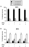
Proliferation of T. gondii in neurons 24 h (A) and 48 h (B) after infection. After 7 days of cultivation neurons were treated with the cytokines indicated for 12 h. After this, RH toxoplasms were added at an MOI of 1. At 24 and 48 h after infection neurons were fixed with 4% paraformaldehyde, and T. gondii was stained immunohistochemically. The number of toxoplasms per PV was determined microscopically for 100 infected neurons per group; the data are means ± standard deviations based on three wells per group. Similar data were obtained in two repeat experiments.
FIG. 3
Cytokine and chemokine production by neurons. Neurons were treated as indicated with IFN-γ and/or TNF for 12 h before RH toxoplasms were added at an MOI of 1. At 48 h after infection the supernatant was harvested, and IL-1β, IL-6, IL-10, GM-CSF, TGF-β1, TGF-β2, MIP-1α, and MIP-1β contents were determined by enzyme-linked immunosorbent assays. IL-10 and GM-CSF were not detected (data not shown). The data are means ± standard deviations based on three wells per group. A second experiment yielded comparable results.
Similar articles
- Cytokine responses induced by Toxoplasma gondii in astrocytes and microglial cells.
Fischer HG, Nitzgen B, Reichmann G, Hadding U. Fischer HG, et al. Eur J Immunol. 1997 Jun;27(6):1539-48. doi: 10.1002/eji.1830270633. Eur J Immunol. 1997. PMID: 9209508 - Infection and replication of human cytomegalovirus in bone marrow stromal cells: effects on the production of IL-6, MIP-1alpha, and TGF-beta1.
Taichman RS, Nassiri MR, Reilly MJ, Ptak RG, Emerson SG, Drach JC. Taichman RS, et al. Bone Marrow Transplant. 1997 Mar;19(5):471-80. doi: 10.1038/sj.bmt.1700685. Bone Marrow Transplant. 1997. PMID: 9052914 - Cytokine expression in the rat adrenal cortex.
Judd AM. Judd AM. Horm Metab Res. 1998 Jun-Jul;30(6-7):404-10. doi: 10.1055/s-2007-978905. Horm Metab Res. 1998. PMID: 9694570 Review. - Interaction between Toxoplasma gondii and enterocyte.
Bout D, Moretto M, Dimier-Poisson I, Gatel DB. Bout D, et al. Immunobiology. 1999 Dec;201(2):225-8. doi: 10.1016/S0171-2985(99)80062-X. Immunobiology. 1999. PMID: 10631571 Review.
Cited by
- Influence of the Host and Parasite Strain on the Immune Response During Toxoplasma Infection.
Mukhopadhyay D, Arranz-Solís D, Saeij JPJ. Mukhopadhyay D, et al. Front Cell Infect Microbiol. 2020 Oct 15;10:580425. doi: 10.3389/fcimb.2020.580425. eCollection 2020. Front Cell Infect Microbiol. 2020. PMID: 33178630 Free PMC article. Review. - Human Neural Stem Cell Systems to Explore Pathogen-Related Neurodevelopmental and Neurodegenerative Disorders.
Baggiani M, Dell'Anno MT, Pistello M, Conti L, Onorati M. Baggiani M, et al. Cells. 2020 Aug 12;9(8):1893. doi: 10.3390/cells9081893. Cells. 2020. PMID: 32806773 Free PMC article. Review. - Toxoplasma on the brain: understanding host-pathogen interactions in chronic CNS infection.
Kamerkar S, Davis PH. Kamerkar S, et al. J Parasitol Res. 2012;2012:589295. doi: 10.1155/2012/589295. Epub 2012 Mar 22. J Parasitol Res. 2012. PMID: 22545203 Free PMC article. - Comprehensive Overview of _Toxoplasma gondii_-Induced and Associated Diseases.
Daher D, Shaghlil A, Sobh E, Hamie M, Hassan ME, Moumneh MB, Itani S, El Hajj R, Tawk L, El Sabban M, El Hajj H. Daher D, et al. Pathogens. 2021 Oct 20;10(11):1351. doi: 10.3390/pathogens10111351. Pathogens. 2021. PMID: 34832507 Free PMC article. Review. - Toxoplasma gondii infection damages the perineuronal nets in a murine model.
Meurer YDSR, Brito RMM, da Silva VP, Andade JMA, Linhares SSG, Pereira Junior A, de Andrade-Neto VF, de Sá AL, Oliveira CBS. Meurer YDSR, et al. Mem Inst Oswaldo Cruz. 2020 Sep 11;115:e200007. doi: 10.1590/0074-02760200007. eCollection 2020. Mem Inst Oswaldo Cruz. 2020. PMID: 32935749 Free PMC article.
References
- Asensio V C, Campbell I L. Chemokines in the CNS: plurifunctional mediators in diverse states. Trends Neurosci. 1999;22:504–512. - PubMed
- Chao C C, Anderson W R, Hu S, Gekker G, Martella A, Peterson P K. Activated microglia inhibit multiplication of Toxoplasma gondii via a nitric oxide mechanism. Clin Immunol Immunopathol. 1993;67:178–183. - PubMed
- Chao C C, Hu S, Gekker G, Novick W J, Jr, Remington J S, Peterson P K. Effects of cytokines on multiplication of Toxoplasma gondii in microglial cells. J Immunol. 1993;150:3404–3410. - PubMed
- Constam D B, Schmid P, Aguzzi A, Schachner M, Fontana A. Transient production of TGF-beta 2 by postnatal cerebellar neurons and its effect on neuroblast proliferation. Eur J Neurosci. 1994;6:766–778. - PubMed
Publication types
MeSH terms
Substances
LinkOut - more resources
Full Text Sources


