SP600125, an anthrapyrazolone inhibitor of Jun N-terminal kinase - PubMed (original) (raw)
. 2001 Nov 20;98(24):13681-6.
doi: 10.1073/pnas.251194298.
D T Sasaki, B W Murray, E C O'Leary, S T Sakata, W Xu, J C Leisten, A Motiwala, S Pierce, Y Satoh, S S Bhagwat, A M Manning, D W Anderson
Affiliations
- PMID: 11717429
- PMCID: PMC61101
- DOI: 10.1073/pnas.251194298
SP600125, an anthrapyrazolone inhibitor of Jun N-terminal kinase
B L Bennett et al. Proc Natl Acad Sci U S A. 2001.
Abstract
Jun N-terminal kinase (JNK) is a stress-activated protein kinase that can be induced by inflammatory cytokines, bacterial endotoxin, osmotic shock, UV radiation, and hypoxia. We report the identification of an anthrapyrazolone series with significant inhibition of JNK1, -2, and -3 (K(i) = 0.19 microM). SP600125 is a reversible ATP-competitive inhibitor with >20-fold selectivity vs. a range of kinases and enzymes tested. In cells, SP600125 dose dependently inhibited the phosphorylation of c-Jun, the expression of inflammatory genes COX-2, IL-2, IFN-gamma, TNF-alpha, and prevented the activation and differentiation of primary human CD4 cell cultures. In animal studies, SP600125 blocked (bacterial) lipopolysaccharide-induced expression of tumor necrosis factor-alpha and inhibited anti-CD3-induced apoptosis of CD4(+) CD8(+) thymocytes. Our study supports targeting JNK as an important strategy in inflammatory disease, apoptotic cell death, and cancer.
Figures
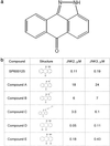
Figure 1
Characterization of SP600125. (a) Chemical structure. (b) Structure-activity relationship study of anthrapyrazolones (IC50 values).
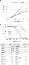
Figure 2
Inhibitory profile of SP600125. (a) SP600125 is an ATP-competitive inhibitor of JNK2 with a _K_i value of 0.19 ± 0.06 μM. At fixed concentrations of JNK2 (9 nM) and c-Jun (2 μM), both ATP and SP600125 concentrations were varied; (□) 0 nM, (○) 50 nM, (▵) 100 nM, and (◊) 300 nM SP600125. The _K_i value was derived from a nonlinear least-squares fit of the data to the kinetic equation for competitive inhibition. (b) SP600125 is a selective inhibitor of JNK. The inhibition profiles of SP600125 vs. related MAP kinases (ERK2 and p38–2) and an unrelated serine threonine kinase (PKA) were performed at _K_m levels of substrates. (c) Kinase selectivity data for SP600125.
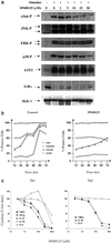
Figure 3
Activity in T cells. (a) Jurkat T cells were resuspended in growth medium and stimulated with phorbol 12-myristate 13-acetate (PMA; 50 ng/ml), anti-CD3 (0.5 μg/ml), and anti-CD28 (2 μg/ml) for 30 min. SP600125 was added as a 10-min pretreatment at the concentrations indicated. The same cell lysates were examined by using different Abs targeting mitogen-activated protein kinase and NF-κB signaling pathways. Levels of nonphosphorylated IKK-1 were used as an indication of equal loading. (b) Inhibition of CD4+ cell activation and differentiation. Th0 cells isolated from either human cord or peripheral blood were cultured with anti-CD3/antiCD28, IL-12, anti-IL-4 in the absence or presence of SP600125 (30 μM). Culture aliquots were analyzed at different times for the expression of cell surface markers CD4, CD45RO, CXCR4, and CCR5. (c) CD4+ cells were differentiated into either Th1 or Th2 cultures over 5 days by using polarizing conditions before stimulating with anti-CD3/anti-CD28 in the presence of increasing concentrations of SP600125 for 15 h in triplicate. Culture supernatants were analyzed by simultaneous analyte reagent technology multiplex cytokine bead array. Data shown represent one of the duplicate experiments. Results are expressed as the mean of triplicate samples.
Figure 4
Inhibitory activity in primary human monocytes. (a) Human PBMCs isolated from peripheral blood were stimulated with LPS (100 ng/ml) for 4 h in the presence of increasing concentrations of SP600125. Cell lysates were prepared and analyzed by using multiplex mRNA detection (Systems Integration Drug Discovery Company). Results are expressed as the mean of triplicate determinations. (b) Primary human monocytes were isolated by positive selection (CD14) and stimulated with LPS (100 ng/ml) in the presence of increasing concentrations of SP600125. After 15 h, culture supernatants were analyzed for expression of IL-1β, -8, and TNF-α with an ELISA. Values are mean and SD for quadruplicate samples.
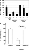
Figure 5
In vivo activity of SP600125. (a) CD-1 mice were dosed with SP600125 i.v. 15 min before injection with LPS or per os 30 min before injection with LPS. At 90 min, a blood sample was recovered, and the serum was obtained. Samples were analyzed for mouse TNF-α by using an ELISA (BioSource). Results are expressed as the mean and standard error with 4 animals per compound treatment group and 6 animals in vehicle control group. Asterisk (*) indicates P ≤ 0.05. DEX, dexamethasone 21-acetate. (b) Apoptosis in murine thymocytes. Thymocytes were isolated from C57BL/6 mice 48 h after injection with anti-CD3 (50 μg) i.p. and analyzed for CD4 and CD8 by flow cytometry. Control animals received no anti-CD3 or vehicle. SP600125 was administered at 0, 12, 24, and 36 h, 15 mg/kg s.c. Anti-CD3 administration was at a single time immediately after the first dosing of SP600125. Data for double-positive (CD4+ CD8+ ) cells is shown expressed as the mean percentage of total thymocytes (n = 4 per group).
Similar articles
- c-Jun N-terminal kinase is required for metalloproteinase expression and joint destruction in inflammatory arthritis.
Han Z, Boyle DL, Chang L, Bennett B, Karin M, Yang L, Manning AM, Firestein GS. Han Z, et al. J Clin Invest. 2001 Jul;108(1):73-81. doi: 10.1172/JCI12466. J Clin Invest. 2001. PMID: 11435459 Free PMC article. - Differential requirement for c-Jun NH2-terminal kinase in TNFalpha- and Fas-mediated apoptosis in hepatocytes.
Schwabe RF, Uchinami H, Qian T, Bennett BL, Lemasters JJ, Brenner DA. Schwabe RF, et al. FASEB J. 2004 Apr;18(6):720-2. doi: 10.1096/fj.03-0771fje. Epub 2004 Feb 6. FASEB J. 2004. PMID: 14766793 - SP600125, an inhibitor of c-jun N-terminal kinase, activates CREB by a p38 MAPK-mediated pathway.
Vaishnav D, Jambal P, Reusch JE, Pugazhenthi S. Vaishnav D, et al. Biochem Biophys Res Commun. 2003 Aug 8;307(4):855-60. doi: 10.1016/s0006-291x(03)01287-7. Biochem Biophys Res Commun. 2003. PMID: 12878189 - Targeting the JNK MAPK cascade for inhibition: basic science and therapeutic potential.
Bogoyevitch MA, Boehm I, Oakley A, Ketterman AJ, Barr RK. Bogoyevitch MA, et al. Biochim Biophys Acta. 2004 Mar 11;1697(1-2):89-101. doi: 10.1016/j.bbapap.2003.11.016. Biochim Biophys Acta. 2004. PMID: 15023353 Review.
Cited by
- Hyperosmotic stress activates the expression of members of the miR-15/107 family and induces downregulation of anti-apoptotic genes in rat liver.
Santosa D, Castoldi M, Paluschinski M, Sommerfeld A, Häussinger D. Santosa D, et al. Sci Rep. 2015 Jul 21;5:12292. doi: 10.1038/srep12292. Sci Rep. 2015. PMID: 26195352 Free PMC article. - Ethacrynic acid and a derivative enhance apoptosis in arsenic trioxide-treated myeloid leukemia and lymphoma cells: the role of glutathione S-transferase p1-1.
Wang R, Liu C, Xia L, Zhao G, Gabrilove J, Waxman S, Jing Y. Wang R, et al. Clin Cancer Res. 2012 Dec 15;18(24):6690-701. doi: 10.1158/1078-0432.CCR-12-0770. Epub 2012 Oct 18. Clin Cancer Res. 2012. PMID: 23082001 Free PMC article. - Mechanisms of Edible Bird's Nest Extract-Induced Proliferation of Human Adipose-Derived Stem Cells.
Roh KB, Lee J, Kim YS, Park J, Kim JH, Lee J, Park D. Roh KB, et al. Evid Based Complement Alternat Med. 2012;2012:797520. doi: 10.1155/2012/797520. Epub 2011 Nov 1. Evid Based Complement Alternat Med. 2012. PMID: 22110547 Free PMC article. - Minimally modified LDL upregulates endothelin type A receptors in rat coronary arterial smooth muscle cells.
Li J, Cao L, Xu CB, Wang JJ, Cao YX. Li J, et al. Mediators Inflamm. 2013;2013:656570. doi: 10.1155/2013/656570. Epub 2013 Jun 19. Mediators Inflamm. 2013. PMID: 23861561 Free PMC article. - Activation of c-Jun N-terminal kinase (JNK) during mitosis in retinal progenitor cells.
Ribas VT, Gonçalves BS, Linden R, Chiarini LB. Ribas VT, et al. PLoS One. 2012;7(4):e34483. doi: 10.1371/journal.pone.0034483. Epub 2012 Apr 4. PLoS One. 2012. PMID: 22496813 Free PMC article.
References
- Derijard B, Hibi M, Wu I-H, Barrett T, Su B, Deng T, Karin M, Davis R J. Cell. 1994;76:1025–1037. - PubMed
- Kallunki T, Su B, Tsigelny I, Sluss H, Derijard B, Moore G, Davis R, Karin M. Genes Dev. 1994;8:2996–3007. - PubMed
- Karin M. J Biol Chem. 1995;270:16483–16486. - PubMed
- Ip Y T, Davis R J. Curr Opin Cell Biol. 1998;10:205–219. - PubMed
- Jain J, Valge-Archer V E, Rao A. J Immunol. 1992;148:1240–1250. - PubMed
MeSH terms
Substances
LinkOut - more resources
Full Text Sources
Other Literature Sources
Molecular Biology Databases
Research Materials
Miscellaneous


