International Union of Pharmacology. XXV. Nomenclature and classification of adenosine receptors - PubMed (original) (raw)
Review
International Union of Pharmacology. XXV. Nomenclature and classification of adenosine receptors
B B Fredholm et al. Pharmacol Rev. 2001 Dec.
Abstract
Four adenosine receptors have been cloned and characterized from several mammalian species. The receptors are named adenosine A(1), A(2A), A(2B), and A(3). The A(2A) and A(2B) receptors preferably interact with members of the G(s) family of G proteins and the A(1) and A(3) receptors with G(i/o) proteins. However, other G protein interactions have also been described. Adenosine is the preferred endogenous agonist at all these receptors, but inosine can also activate the A(3) receptor. The levels of adenosine seen under basal conditions are sufficient to cause some activation of all the receptors, at least where they are abundantly expressed. Adenosine levels during, e.g., ischemia can activate all receptors even when expressed in low abundance. Accordingly, experiments with receptor antagonists and mice with targeted disruption of adenosine A(1), A(2A), and A(3) expression reveal roles for these receptors under physiological and particularly pathophysiological conditions. There are pharmacological tools that can be used to classify A(1), A(2A), and A(3) receptors but few drugs that interact selectively with A(2B) receptors. Testable models of the interaction of these drugs with their receptors have been generated by site-directed mutagenesis and homology-based modelling. Both agonists and antagonists are being developed as potential drugs.
Figures
FIG. 1.
Dendrogram showing sequence similarity between cloned adenosine receptors. The figure is slightly redrawn from that available at
http://www.gpcr.org/7tm/seq/007\_001/001\_007\_001.TREE.html
. This phylogenetic tree was automatically calculated by WHAT IF based on a neighbor-joining algorithm.
FIG. 2.
Protein backbone representations of the structures of bacteriorhodopsin (A) and rhodopsin (B) as retrieved from the Brookhaven Protein Data Bank (1C3W and 1F88, respectively).
FIG. 3.
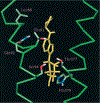
Computer modeling of CPA (yellow), human A1 adenosine receptor interactions. Only helices III (left) and VII (right) are shown (both in green) with relevant residues (half-bond colors).
FIG. 4.
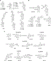
Structures of reference compounds used to classify adenosine receptors. Panel A, structure of some non-selective and A1 receptor-selective adenosine analogs; panel B, structure of adenosine analogs used to classify A2A and A3 receptors; panel C, structure of selected adenosine receptor antagonists.
Similar articles
- International Union of Basic and Clinical Pharmacology. LXXXI. Nomenclature and classification of adenosine receptors--an update.
Fredholm BB, IJzerman AP, Jacobson KA, Linden J, Müller CE. Fredholm BB, et al. Pharmacol Rev. 2011 Mar;63(1):1-34. doi: 10.1124/pr.110.003285. Epub 2011 Feb 8. Pharmacol Rev. 2011. PMID: 21303899 Free PMC article. Review. - Medicinal chemistry and pharmacology of A2B adenosine receptors.
Volpini R, Costanzi S, Vittori S, Cristalli G, Klotz KN. Volpini R, et al. Curr Top Med Chem. 2003;3(4):427-43. doi: 10.2174/1568026033392264. Curr Top Med Chem. 2003. PMID: 12570760 - Introduction to adenosine receptors as therapeutic targets.
Jacobson KA. Jacobson KA. Handb Exp Pharmacol. 2009;(193):1-24. doi: 10.1007/978-3-540-89615-9_1. Handb Exp Pharmacol. 2009. PMID: 19639277 Free PMC article. Review. - Nomenclature and classification of purinoceptors.
Fredholm BB, Abbracchio MP, Burnstock G, Daly JW, Harden TK, Jacobson KA, Leff P, Williams M. Fredholm BB, et al. Pharmacol Rev. 1994 Jun;46(2):143-56. Pharmacol Rev. 1994. PMID: 7938164 Free PMC article. Review. No abstract available. - Dominance of G(s) in doubly G(s)/G(i)-coupled chimaeric A(1)/A(2A) adenosine receptors in HEK-293 cells.
Tucker AL, Jia LG, Holeton D, Taylor AJ, Linden J. Tucker AL, et al. Biochem J. 2000 Nov 15;352 Pt 1(Pt 1):203-10. Biochem J. 2000. PMID: 11062074 Free PMC article.
Cited by
- Insulin-increased L-arginine transport requires A(2A) adenosine receptors activation in human umbilical vein endothelium.
Guzmán-Gutiérrez E, Westermeier F, Salomón C, González M, Pardo F, Leiva A, Sobrevia L. Guzmán-Gutiérrez E, et al. PLoS One. 2012;7(7):e41705. doi: 10.1371/journal.pone.0041705. Epub 2012 Jul 23. PLoS One. 2012. PMID: 22844517 Free PMC article. - Exploration of the link between gut microbiota and purinergic signalling.
Li M, Liu B, Li R, Yang P, Leng P, Huang Y. Li M, et al. Purinergic Signal. 2023 Mar;19(1):315-327. doi: 10.1007/s11302-022-09891-1. Epub 2022 Sep 19. Purinergic Signal. 2023. PMID: 36121551 Free PMC article. Review. - Contribution of pannexin1 to experimental autoimmune encephalomyelitis.
Lutz SE, González-Fernández E, Ventura JC, Pérez-Samartín A, Tarassishin L, Negoro H, Patel NK, Suadicani SO, Lee SC, Matute C, Scemes E. Lutz SE, et al. PLoS One. 2013 Jun 20;8(6):e66657. doi: 10.1371/journal.pone.0066657. Print 2013. PLoS One. 2013. PMID: 23885286 Free PMC article. - Adenosine 2A receptors in acute kidney injury.
Vincent IS, Okusa MD. Vincent IS, et al. Acta Physiol (Oxf). 2015 Jul;214(3):303-10. doi: 10.1111/apha.12508. Epub 2015 May 19. Acta Physiol (Oxf). 2015. PMID: 25877257 Free PMC article. Review. - Inosine: A bioactive metabolite with multimodal actions in human diseases.
Kim IS, Jo EK. Kim IS, et al. Front Pharmacol. 2022 Nov 16;13:1043970. doi: 10.3389/fphar.2022.1043970. eCollection 2022. Front Pharmacol. 2022. PMID: 36467085 Free PMC article. Review.
References
- Abbracchio MP, Ceruti S, Brambilla R, Franceschi C, Malorni W, Jacobson KA, von Lubitz DK, and Cattabeni F (1997) Modulation of apoptosis by adenosine in the central nervous system: a possible role for the A3 receptor. Pathophysiological significance and therapeutic implications for neurodegenerative disorders. Ann NY Acad Sci 825:11–22. - PMC - PubMed
- Ådén U, Leverin A-L, Hagberg H, and Fredholm BB (2001) Adenosine A1 receptor agonism in the immature rat brain and heart. Eur J Pharmacol 426:185–192. - PubMed
- Ådén U, Herlenius E, Tang L-Q, and Fredholm BB (2000) Maternal caffeine intake has minor effects on adenosine receptor ontogeny in the rat brain. Pediat Res 48:177–183. - PubMed
- Akbar M, Okajima F, Tomura H, Shimegi S, and Kondo Y (1994) A single species of A1 adenosine receptor expressed in Chinese hamster ovary cells not only inhibits cAMP accumulation but also stimulates phospholipase C and arachidonate release. Mol Pharmacol 45:1036–1042. - PubMed
- Altiok N, Balmforth AJ, and Fredholm BB (1992) Adenosine receptor-induced cAMP changes in D384 astrocytoma cells and the effect of bradykinin thereon. Acta Physiol Scand 144:55–63. - PubMed
Publication types
MeSH terms
Substances
LinkOut - more resources
Full Text Sources
Other Literature Sources
Molecular Biology Databases


