PTP and LTP at a hippocampal mossy fiber-interneuron synapse - PubMed (original) (raw)
PTP and LTP at a hippocampal mossy fiber-interneuron synapse
H Alle et al. Proc Natl Acad Sci U S A. 2001.
Abstract
The mossy fiber-CA3 pyramidal neuron synapse is a main component of the hippocampal trisynaptic circuitry. Recent studies, however, suggested that inhibitory interneurons are the major targets of the mossy fiber system. To study the regulation of mossy fiber-interneuron excitation, we examined unitary and compound excitatory postsynaptic currents in dentate gyrus basket cells, evoked by paired recording between granule and basket cells or extracellular stimulation of mossy fiber collaterals. The application of an associative high-frequency stimulation paradigm induced posttetanic potentiation (PTP) followed by homosynaptic long-term potentiation (LTP). Analysis of numbers of failures, coefficient of variation, and paired-pulse modulation indicated that both PTP and LTP were expressed presynaptically. The Ca(2+) chelator 1,2-bis(2-aminophenoxy)ethane-N,N,N',N'-tetraacetic acid (BAPTA) did not affect PTP or LTP at a concentration of 10 mM but attenuated LTP at a concentration of 30 mM. Both forskolin, an adenylyl cyclase activator, and phorbolester diacetate, a protein kinase C stimulator, lead to a long-lasting increase in excitatory postsynaptic current amplitude. H-89, a protein kinase A inhibitor, and bisindolylmaleimide, a protein kinase C antagonist, reduced PTP, whereas only bisindolylmaleimide reduced LTP. These results may suggest a differential contribution of protein kinase A and C pathways to mossy fiber-interneuron plasticity. Interneuron PTP and LTP may provide mechanisms to maintain the balance between synaptic excitation of interneurons and that of principal neurons in the dentate gyrus-CA3 network.
Figures
Figure 1
PTP and LTP of unitary EPSCs at the mossy fiber-basket cell synapse. (A) Schematic illustration of the paired recording configuration. GC, granule cell; BC, basket cell; GCL, granule cell layer; CA3, CA3 subfield; MF, mossy fiber. The figure was adapted from ref. . (B) An aHFS induces both PTP and LTP. Single presynaptic APs are shown on top, and average EPSCs (from 64, 10, and 100 single traces in the time intervals before the first aHFS, directly after the first aHFS, and 10–15 min after the third aHFS, respectively) are depicted below. Boxes indicate details of the aHFSs. (C and D) PTP and LTP of EPSCs induced by a single aHFS. Unitary EPSC peak amplitudes from paired recordings plotted against time. Data from a single paired recording are shown in_C_, and the summary graph for eight pairs is shown in_D_ (with EPSC amplitudes normalized to the control value). The aHFS was applied at t = 0, as indicated by the arrow. (E and F) PTP and LTP of EPSCs induced by triple aHFS. Three single aHFS protocols were separated by 1.5-min intervals. Data from a single paired recording are shown in E, and the summary graph for three pairs is shown in F (●). For comparison, the effect of triple nHFS with the postsynaptic cell held in the voltage-clamp configuration at −70 mV during induction is depicted also (○, three pairs). The data shown in B and E were taken from the same experiment. Curves in_D_ and F represent exponential functions fitted to the data points with time constants of 1.7 (D), 3.6 (F, aHFS), and 2.4 min (F, nHFS). The recording temperature was 34 ± 2°C in these and all subsequent experiments.
Figure 2
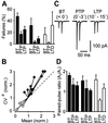
Presynaptic locus of PTP and LTP of unitary EPSCs at the mossy fiber-basket cell synapse. (A) Bar graph of the percentage of failures during BT, PTP, and LTP/LTD (with time intervals identical to those specified in Fig. 1). The mean effects of single aHFS (filled bars, left), triple aHFS (filled bars, center), and triple nHFS (open bars, right). (B) CV analysis of long-term changes. The CV−2 of the EPSC amplitude in the late phase after induction was plotted against mean, both normalized to their respective control values before HFS. The effects of single aHFS (●), triple aHFS (♦), and triple nHFS (○) are shown. The location of the data points with respect to the identity line (dashed) was consistent with presynaptic changes in all cases. (C) Average unitary EPSCs (from 25, 15, and 25 single traces during BT, PTP, and LTP phases, respectively; single aHFS protocol) evoked by pairs of two APs, separated by 20-ms intervals. (D) Paired-pulse ratios for BT, PTP, and LTP/LTD periods. The mean effects of single aHFS (filled bars, left), triple aHFS (filled bars, center), and triple nHFS (open bars, right) are shown. The paired-pulse ratio was determined as_A_2/_A_1 from average trace.
Figure 3
The aHFS-induced LTP of compound EPSCs shows weak sensitivity to the Ca2+ chelator BAPTA. (A) Compound EPSCs are similar to unitary EPSCs with respect to PTP, LTP after aHFS, and DCG-4 sensitivity. Aa, average compound EPSCs in BT and LTP phases (stimulus artifacts blanked); Ab, corresponding EPSC peak amplitude values plotted against time. (B) Bar graph of compound EPSC amplitude (filled) and unitary EPSC amplitude (open) in the presence of 0.5–1 μM DCG-4 relative to respective control values. (C and D) Effects of single aHFS on compound EPSC amplitude with 0.1 mM intracellular EGTA (●; 11 cells), 10 mM intracellular BAPTA (C, ○; six cells), and 30 mM BAPTA (D, ○; eight cells). Amplitudes were normalized to the mean EPSC peak amplitude before aHFS. The aHFS was applied at t = 0, as indicated by the arrow. (E and F) Cumulative distributions of the extent of aHFS-induced PTP (E) and LTP (F) of compound EPSCs in individual experiments in 0.1 mM internal EGTA, 10 mM BAPTA, and 30 mM BAPTA. Paired recording data (with 0.2 mM internal EGTA) are shown superimposed.
Figure 4
Involvement of PKA and PKC pathways in aHFS-induced PTP and LTP of compound EPSCs. (A) Potentiation of compound EPSC amplitude (normalized to BT period) by bath application of 50 μM forskolin (○; six cells) and 1 μM PDA (●; five cells; horizontal bars indicate application times). (B) CV analysis of forskolin- (○) and PDA- (●) induced potentiation. The CV−2 of the EPSC amplitude 10–15 min (forskolin, two cells), 15–20 min (forskolin, four cells; PDA, one cell), and 20–25 min (PDA, four cells) after the onset of application was plotted against mean, both normalized to their respective control values. Location of the data points with respect to the identity line (dashed) suggests presynaptic changes. (C and D) Effects of single aHFS on compound EPSC amplitude after preincubation with 10 μM H-89 (C, seven cells) and 1 μM bisindolylmaleimide (D, 11 cells). The aHFS was applied at_t_ = 0, as indicated by the arrow. Curves in_C_ and D represent exponential function fitted to the control data points (0.1 mM EGTA) in Fig. 3_C_ and D (●; time constant 1.7 min). (E and F) Cumulative distributions of the extent of aHFS-induced PTP (E) and LTP (F) of compound EPSCs in individual experiments in control conditions (same experiments as shown in Fig. 3_C_), after preincubation with 10 μM H-89 (C), and one after preincubation with 1 μM bisindolylmaleimide (D).
Similar articles
- Critical involvement of postsynaptic protein kinase activation in long-term potentiation at hippocampal mossy fiber synapses on CA3 interneurons.
Galván EJ, Cosgrove KE, Mauna JC, Card JP, Thiels E, Meriney SD, Barrionuevo G. Galván EJ, et al. J Neurosci. 2010 Feb 24;30(8):2844-55. doi: 10.1523/JNEUROSCI.5269-09.2010. J Neurosci. 2010. PMID: 20181582 Free PMC article. - Synapse-specific compartmentalization of signaling cascades for LTP induction in CA3 interneurons.
Galván EJ, Pérez-Rosello T, Gómez-Lira G, Lara E, Gutiérrez R, Barrionuevo G. Galván EJ, et al. Neuroscience. 2015 Apr 2;290:332-45. doi: 10.1016/j.neuroscience.2015.01.024. Epub 2015 Jan 28. Neuroscience. 2015. PMID: 25637803 Free PMC article. - Mediation of hippocampal mossy fiber long-term potentiation by presynaptic Ih channels.
Mellor J, Nicoll RA, Schmitz D. Mellor J, et al. Science. 2002 Jan 4;295(5552):143-7. doi: 10.1126/science.1064285. Science. 2002. PMID: 11778053 - Multiple forms of long-term synaptic plasticity at hippocampal mossy fiber synapses on interneurons.
Galván EJ, Cosgrove KE, Barrionuevo G. Galván EJ, et al. Neuropharmacology. 2011 Apr;60(5):740-7. doi: 10.1016/j.neuropharm.2010.11.008. Epub 2010 Nov 18. Neuropharmacology. 2011. PMID: 21093459 Free PMC article. Review. - TwoB or not twoB: differential transmission at glutamatergic mossy fiber-interneuron synapses in the hippocampus.
Bischofberger J, Jonas P. Bischofberger J, et al. Trends Neurosci. 2002 Dec;25(12):600-3. doi: 10.1016/s0166-2236(02)02259-2. Trends Neurosci. 2002. PMID: 12446120 Review.
Cited by
- Cell-specific synaptic plasticity induced by network oscillations.
Zarnadze S, Bäuerle P, Santos-Torres J, Böhm C, Schmitz D, Geiger JR, Dugladze T, Gloveli T. Zarnadze S, et al. Elife. 2016 May 24;5:e14912. doi: 10.7554/eLife.14912. Elife. 2016. PMID: 27218453 Free PMC article. - Associative plasticity at excitatory synapses facilitates recruitment of fast-spiking interneurons in the dentate gyrus.
Sambandan S, Sauer JF, Vida I, Bartos M. Sambandan S, et al. J Neurosci. 2010 Sep 1;30(35):11826-37. doi: 10.1523/JNEUROSCI.2012-10.2010. J Neurosci. 2010. PMID: 20810902 Free PMC article. - Phosphorylation of synapsin domain A is required for post-tetanic potentiation.
Fiumara F, Milanese C, Corradi A, Giovedì S, Leitinger G, Menegon A, Montarolo PG, Benfenati F, Ghirardi M. Fiumara F, et al. J Cell Sci. 2007 Sep 15;120(Pt 18):3228-37. doi: 10.1242/jcs.012005. Epub 2007 Aug 28. J Cell Sci. 2007. PMID: 17726061 Free PMC article. - Role of ionotropic glutamate receptors in long-term potentiation in rat hippocampal CA1 oriens-lacunosum moleculare interneurons.
Oren I, Nissen W, Kullmann DM, Somogyi P, Lamsa KP. Oren I, et al. J Neurosci. 2009 Jan 28;29(4):939-50. doi: 10.1523/JNEUROSCI.3251-08.2009. J Neurosci. 2009. PMID: 19176803 Free PMC article. - Critical involvement of postsynaptic protein kinase activation in long-term potentiation at hippocampal mossy fiber synapses on CA3 interneurons.
Galván EJ, Cosgrove KE, Mauna JC, Card JP, Thiels E, Meriney SD, Barrionuevo G. Galván EJ, et al. J Neurosci. 2010 Feb 24;30(8):2844-55. doi: 10.1523/JNEUROSCI.5269-09.2010. J Neurosci. 2010. PMID: 20181582 Free PMC article.
References
- Freund T F, Buzsáki G. Hippocampus. 1996;6:347–470. - PubMed
- Cobb S R, Buhl E H, Halasy K, Paulsen O, Somogyi P. Nature (London) 1995;378:75–78. - PubMed
- Davies C H, Starkey S J, Pozza M F, Collingridge G L. Nature (London) 1991;349:609–611. - PubMed
Publication types
MeSH terms
Substances
LinkOut - more resources
Full Text Sources
Miscellaneous


