Adult neurogenesis in mammals: an identity crisis - PubMed (original) (raw)
Review
Adult neurogenesis in mammals: an identity crisis
Pasko Rakic. J Neurosci. 2002.
No abstract available
Figures
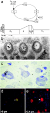
Fig. 1.
a, Graphic representation of the level of DNA during different phases of cell cycle (G 1 and_G2_, gap phases; D, mitotic division; S, synthesis phase). The synthesis of DNA can be detected by the incorporation of 3H-dT or its analog BrdU. The method of 3H-dT incorporation after a single injection of the isotope is stoichiometric, and thus allows distinction between doubling of DNA content during the S phase, signifying mitotic division, and the light label of <50% of the maximum grain counts. b, The Purkinje cells in the_middle_ and on the left can be considered as heavily labeled, whereas the five grains over the cell nucleus on the right may be caused by the variety of biological and technological factors, some of which are discussed in the text. Importantly, if the animal were injected with 10, instead of a single3H-dT dose, this cell may be as heavily labeled as the one in the middle and could be falsely interpreted as divided.c, Interference (Nomarski) contrast microphotograph of a heavily labeled satellite glial cell in the frontal lobe of a monkey found 35 d after a single injection of 3H-dT (10 mCi/kg). d, Image of a cell in the adult macaque monkey prefrontal cortex that appears to be double-labeled for BrdU and NeuN, but was proven to belong to two closely apposed single-labeled cells by the use of z-series analyses on a confocal microscope. An NeuN-positive neuron (red) appeared to be colabeled with BrdU (green) at +0 level (arrow in_d_), but examination of a different focal plane (+2.8 μm level) reveals that the NeuN-positive nucleus and nucleolus actually belong to a neuron that is not BrdU-labeled (arrowhead in e). The BrdU-labeled nucleus apparently belongs to adult-generated satellite glia, which in the cerebral cortex typically associate with neuronal perikarya as seen also in c. Astrocytes in d and_e_ are indicated by GFAP immunoreactivity (blue). For further information, see Kornack and Rakic (2001b) and Rakic (2002).
Fig. 2.
Exogenous 3H-dT (red dots) or BrdU (blue squares) compete with the natural endogenous H-dT for incorporation into nuclear DNA.a, During S phase of cell cycle, incorporation occurs predominately into new stands of DNA, and provided that the cell stops dividing, significant presence of these marks indicates the time of final cell division. b, Incorporation of nucleotides occurs at a slower rate as part of DNA turnover or repair (of both strands) or at the higher rate during an abortive cell cycle (predominately in a single strand) leading to recovery or death (for further explanation, see text and Fig. 1).
Similar articles
- Neurogenesis in adult mammals: some progress and problems.
Gould E, Gross CG. Gould E, et al. J Neurosci. 2002 Feb 1;22(3):619-23. doi: 10.1523/JNEUROSCI.22-03-00619.2002. J Neurosci. 2002. PMID: 11826089 Free PMC article. Review. No abstract available. - Continual replacement of newly-generated olfactory neurons in adult rats.
Kato T, Yokouchi K, Fukushima N, Kawagishi K, Li Z, Moriizumi T. Kato T, et al. Neurosci Lett. 2001 Jul 6;307(1):17-20. doi: 10.1016/s0304-3940(01)01914-0. Neurosci Lett. 2001. PMID: 11516564 - The cell biology of neurogenesis.
Götz M, Huttner WB. Götz M, et al. Nat Rev Mol Cell Biol. 2005 Oct;6(10):777-88. doi: 10.1038/nrm1739. Nat Rev Mol Cell Biol. 2005. PMID: 16314867 Review. - Life-long stability of neurons: a century of research on neurogenesis, neuronal death and neuron quantification in adult CNS.
Turlejski K, Djavadian R. Turlejski K, et al. Prog Brain Res. 2002;136:39-65. doi: 10.1016/s0079-6123(02)36006-0. Prog Brain Res. 2002. PMID: 12143397 Review. - Cell division and differentiation of central nervous system neurons.
Tixier-Vidal A. Tixier-Vidal A. Ann N Y Acad Sci. 1994 Sep 15;733:56-67. doi: 10.1111/j.1749-6632.1994.tb17256.x. Ann N Y Acad Sci. 1994. PMID: 7978903 Review. No abstract available.
Cited by
- In vivo imaging of juxtaglomerular neuron turnover in the mouse olfactory bulb.
Mizrahi A, Lu J, Irving R, Feng G, Katz LC. Mizrahi A, et al. Proc Natl Acad Sci U S A. 2006 Feb 7;103(6):1912-7. doi: 10.1073/pnas.0506297103. Epub 2006 Jan 30. Proc Natl Acad Sci U S A. 2006. PMID: 16446451 Free PMC article. - Programmed cell death of adult-generated hippocampal neurons is mediated by the proapoptotic gene Bax.
Sun W, Winseck A, Vinsant S, Park OH, Kim H, Oppenheim RW. Sun W, et al. J Neurosci. 2004 Dec 8;24(49):11205-13. doi: 10.1523/JNEUROSCI.1436-04.2004. J Neurosci. 2004. PMID: 15590937 Free PMC article. - Neuron-like differentiation of bone marrow-derived mesenchymal stem cells.
Bae KS, Park JB, Kim HS, Kim DS, Park DJ, Kang SJ. Bae KS, et al. Yonsei Med J. 2011 May;52(3):401-12. doi: 10.3349/ymj.2011.52.3.401. Yonsei Med J. 2011. PMID: 21488182 Free PMC article. - Reactive Neuroblastosis in Huntington's Disease: A Putative Therapeutic Target for Striatal Regeneration in the Adult Brain.
Kandasamy M, Aigner L. Kandasamy M, et al. Front Cell Neurosci. 2018 Mar 9;12:37. doi: 10.3389/fncel.2018.00037. eCollection 2018. Front Cell Neurosci. 2018. PMID: 29593498 Free PMC article. - Running wheel training does not change neurogenesis levels or alter working memory tasks in adult rats.
Acevedo-Triana CA, Rojas MJ, Cardenas FP. Acevedo-Triana CA, et al. PeerJ. 2017 May 9;5:e2976. doi: 10.7717/peerj.2976. eCollection 2017. PeerJ. 2017. PMID: 28503368 Free PMC article.
References
- Anatskaya OV, Vinogradov AE, Kudryavtsev BN. Hepatocyte polyploidy and metabolism/life-history traits: hypotheses testing. J Theor Biol. 1994;168:191–199. - PubMed
- Anderson JD, Gage FH, Weissman IL. Can stem cells cross lineage boundaries. Nat Med. 2001;7:393–395. - PubMed
- Angevine JB., Jr Time of neuron origin in the hippocampal region. An autoradiographic study in the mouse. Exp Neurol [Suppl] 1965;2:1–70. - PubMed
- Brazelton TR, Rossi FM, Keshet GI, Blau HM. From marrow to brain: expression of neuronal phenotypes in adult mice. Science. 2000;290:1775–1779. - PubMed
Publication types
MeSH terms
Substances
LinkOut - more resources
Full Text Sources

