Proteasome inhibition reduces superantigen-mediated T cell activation and the severity of psoriasis in a SCID-hu model - PubMed (original) (raw)
Proteasome inhibition reduces superantigen-mediated T cell activation and the severity of psoriasis in a SCID-hu model
Thomas M Zollner et al. J Clin Invest. 2002 Mar.
Abstract
There is increasing evidence that bacterial superantigens contribute to inflammation and T cell responses in psoriasis. Psoriatic inflammation entails a complex series of inductive and effector processes that require the regulated expression of various proinflammatory genes, many of which require NF-kappa B for maximal trans-activation. PS-519 is a potent and selective proteasome inhibitor based upon the naturally occurring compound lactacystin, which inhibits NF-kappa B activation by blocking the degradation of its inhibitory protein I kappa B. We report that proteasome inhibition by PS-519 reduces superantigen-mediated T cell-activation in vitro and in vivo. Proliferation was inhibited along with the expression of very early (CD69), early (CD25), and late T cell (HLA-DR) activation molecules. Moreover, expression of E-selectin ligands relevant to dermal T cell homing was reduced, as was E-selectin binding in vitro. Finally, PS-519 proved to be therapeutically effective in a SCID-hu xenogeneic psoriasis transplantation model. We conclude that inhibition of the proteasome, e.g., by PS-519, is a promising means to treat T cell-mediated disorders such as psoriasis.
Figures
Figure 1
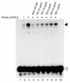
The PHA/IL-2/TGF-β–induced NF-κB DNA complex is suppressed by the proteasome inhibitor PS-519. Human T cells were stimulated with PHA/IL-2/TGF-β for 4 hours. PS-519 (1–10 μg/ml) suppressed NF-κB DNA-binding activity. The NF-κB DNA complex is indicated by a filled arrowhead. The open arrowhead shows the position of the unbound DNA probe. Binding mixture without cell extract was applied on lanes 1 and 10. The antioxidant _N_-acetylcysteine (NAC; 25 mM), which is known to inhibit IκB kinase, served as control (lane 4). The results are representative of three electrophoretic mobility shift assays from three independent donors.
Figure 2
PS-519 inhibits TSST-1–induced T cell proliferation. PBMCs (2 × 10⁶/ml) obtained from five healthy volunteers were stimulated with TSST-1 (100 ng/ml) in the absence or presence of PS-519 (0.25–2.5 μg/ml) for 4 days and thereafter pulsed with 3H-thymidine. Incorporation of 3H-thymidine into DNA was calculated using a liquid scintillation counter. Stimulation index was calculated by the ratio: decays per minute of experimental group/decays per minute of control group. Open symbols represent resting PBMCs, filled symbols TSST-1–stimulated PBMCs. A significant reduction in proliferation was observed starting at 0.25 μg/ml PS-519 (*P < 0.001). Values represent mean ± SD of five healthy donors.
Figure 3
PS-519 inhibits TSST-1–induced expression of T cell activation molecules. PBMCs were stimulated with TSST-1 (100 ng/ml) in the presence or absence of PS-519 (0.25–2.5 μg/ml). CD69+ CD3+ (a), CD25+ CD3+ (b), and HLA-DR+ CD3+ (c) surface expression was measured at days 1, 3, 5, 7, and 9 (days 7 and 9 not shown) by flow cytometry. Appropriate isotype Ig’s served as controls to set gates for positive and negative staining. For CD69 expression, significant reduction was observed on day 1 starting at 1.0 μg/ml (P < 0.05), and on days 3 and 5 starting at 0.5 μg/ml PS-519 (P < 0.001 and P < 0.05, respectively). For CD25 expression, significant reduction was observed on day 3 starting at 1.0 μg/ml (P < 0.001), and on day 5 starting at 0.25 μg/ml PS-519 (P < 0.001). For HLA-DR expression, significant reduction was observed on day 1 at 2.5 μg/ml (P < 0.05), on day 3 starting at 1.0 μg/ml (P < 0.05), and on day 5 starting at 0.25 μg/ml PS-519 (P < 0.001). Data represent means of five experiments ± SD.
Figure 4
PS-519 inhibits TSST-1–induced expression of T cell adhesion molecules. PBMCs were stimulated with TSST-1 (100 ng/ml) in the presence or absence of PS-519 (0.25–2.5 μg/ml). CLA+CD3+ (a) and CD15s+CD3+ (b) surface expression and binding of CD3+ cells to E-selectin (c) was measured at days 1, 3, 5, 7, and 9 (days 1 and 9 not shown) by flow cytometry. Appropriate isotype Ig’s served as controls to set gates for positive and negative staining. Staining in the absence of fusion proteins and in the presence of anti-CD3–FITC together with secondary anti-human IgG-phycoerythrin demonstrated absence of unspecific binding reactivity. For CLA expression, significant reduction was observed on day 3 at 2.5 μg/ml (P < 0.05), and on days 5 and 7 starting at 0.25 μg/ml PS-519 (P < 0.001). For CD15s expression, significant reduction was observed on days 5 and 7 starting at 0.25 μg/ml PS-519 (P < 0.001). For E-selectin binding, significant reduction was observed on day 5 starting at 0.25 μg/ml (P < 0.001), and on day 7 starting at 0.5 μg/ml PS-519 (P < 0.05). Data represent means of five independent experiments ± SD.
Figure 5
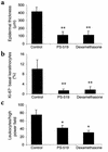
PS-519 suppresses hallmarks of psoriasis in a xenogeneic transplantation model. Grafts from lesional psoriatic skin were transplanted onto SCID mice as outlined in Methods. After 2 weeks, mice were treated with PS-519 (1 mg/kg body weight), dexamethasone (0.2 mg/kg body weight), or vehicle for 4 weeks. Subsequently, epidermal thickness (a), proliferation of basal keratinocytes measured by Ki-67 reactivity (b), and leukocytic infiltration (c) were determined by a blinded investigator. All parameters showed a marked reduction (*P < 0.05, **P < 0.001). Data represent means of four independent experiments ± SD.
Figure 6
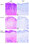
PS-519 suppresses hallmarks of psoriasis in a xenogeneic transplantation model. Grafted skin in PS-519–treated mice (1 mg/kg body weight) showed normalization of epidermal architecture, loss of papillomatosis, and marked reduction of acanthosis (c, hematoxylin-and-eosin stain) as compared with vehicle-treated mice (a, hematoxylin-and-eosin stain). In PS-519–treated (d, Ki-67 stain) as compared with vehicle-treated mice (b, Ki-67 stain), proliferation of basal keratinocytes was markedly reduced. Treatment with dexamethasone (0.2 mg/kg body weight; e and f) was as effective as PS-519 treatment. The sections show one representative of four experiments. ×200.
Figure 7
20S proteasome activity is reduced in PS-519 as compared with vehicle treated mice. Peripheral blood from vehicle- and PS-519–treated mice was drawn 2 hours after the final injection at days 4, 8, 14, and 28. Thereafter, 20S proteasome activity was determined as described above. PS-519–treated animals showed an 85.7% ± 8.6% (mean ± SD) inhibition of the 20S proteasome activity as compared with vehicle-treated mice at day 28 (**P < 0.0001). Already after 4 days, 81.7% ± 1.6% inhibition as compared with controls was achieved (*P < 0.01). The 20S proteasome activity in vehicle-treated mice remained unchanged when measured at 4, 8, and 14 days as compared with day 28 (data not shown). The 20S proteasome activity is given in pmol/s/mg protein. Data represent mean ± SD; n = 6, days 4–14; n = 8, day 28.
Similar articles
- Anti-inflammatory effects of plumbagin are mediated by inhibition of NF-kappaB activation in lymphocytes.
Checker R, Sharma D, Sandur SK, Khanam S, Poduval TB. Checker R, et al. Int Immunopharmacol. 2009 Jul;9(7-8):949-58. doi: 10.1016/j.intimp.2009.03.022. Epub 2009 Apr 15. Int Immunopharmacol. 2009. PMID: 19374955 - Vbeta-restricted T cell adherence to endothelial cells: a mechanism for superantigen-dependent vascular injury.
Brogan PA, Shah V, Klein N, Dillon MJ. Brogan PA, et al. Arthritis Rheum. 2004 Feb;50(2):589-97. doi: 10.1002/art.20021. Arthritis Rheum. 2004. PMID: 14872503 - In vivo T cell response to viral superantigen. Selective migration rather than proliferation.
Le Bon A, Lucas B, Vasseur F, Penit C, Papiernik M. Le Bon A, et al. J Immunol. 1996 Jun 15;156(12):4602-8. J Immunol. 1996. PMID: 8648102 - The proteasome system and proteasome inhibitors in stroke: controlling the inflammatory response.
Di Napoli M, Papa F. Di Napoli M, et al. Curr Opin Investig Drugs. 2003 Nov;4(11):1333-42. Curr Opin Investig Drugs. 2003. PMID: 14758773 Review. - Biologic effects of bacterial superantigens in a xenogeneic transplantation model for psoriasis.
Boehncke WH. Boehncke WH. J Investig Dermatol Symp Proc. 2001 Dec;6(3):231-2. doi: 10.1046/j.0022-202x.2001.00042.x. J Investig Dermatol Symp Proc. 2001. PMID: 11924833 Review.
Cited by
- Proteasome inhibition: a new anti-inflammatory strategy.
Elliott PJ, Zollner TM, Boehncke WH. Elliott PJ, et al. J Mol Med (Berl). 2003 Apr;81(4):235-45. doi: 10.1007/s00109-003-0422-2. Epub 2003 Mar 26. J Mol Med (Berl). 2003. PMID: 12700891 Review. - [Psoriasis SCID-mouse model].
Pfeffer J, Kaufmann R, Boehncke WH. Pfeffer J, et al. Hautarzt. 2006 Jul;57(7):603-9. doi: 10.1007/s00105-005-0990-x. Hautarzt. 2006. PMID: 16028077 Review. German. - Translational Clinical Strategies for the Prevention of Gastrointestinal Tract Graft Versus Host Disease.
Rayasam A, Drobyski WR. Rayasam A, et al. Front Immunol. 2021 Nov 26;12:779076. doi: 10.3389/fimmu.2021.779076. eCollection 2021. Front Immunol. 2021. PMID: 34899738 Free PMC article. Review. - Proteasome inhibition alleviates prolonged moderate compression-induced muscle pathology.
Siu PM, Teng BT, Pei XM, Tam EW. Siu PM, et al. BMC Musculoskelet Disord. 2011 Mar 7;12:58. doi: 10.1186/1471-2474-12-58. BMC Musculoskelet Disord. 2011. PMID: 21385343 Free PMC article. - Recent insights into the immunopathogenesis of psoriasis provide new therapeutic opportunities.
Nickoloff BJ, Nestle FO. Nickoloff BJ, et al. J Clin Invest. 2004 Jun;113(12):1664-75. doi: 10.1172/JCI22147. J Clin Invest. 2004. PMID: 15199399 Free PMC article. Review.
References
- Acha-Orbea, H. 1995. Superantigens and tolerance. In T cell receptors. J.I. Bell, M.J. Owen, and E. Simpson, editors. Oxford University Press. Oxford, United Kingdom. 224–265.
- Zollner TM, Nuber V, Duijvestijn AM, Boehncke WH, Kaufmann R. Superantigens but not mitogens are capable of inducing upregulation of E-selectin ligands on human T lymphocytes. Exp Dermatol. 1997;6:161–166. - PubMed
- Zollner TM, et al. The superantigen exfoliative toxin induces cutaneous lymphocyte-associated antigen expression in peripheral human T lymphocytes. Immunol Lett. 1996;49:111–116. - PubMed
- Paliard X, et al. Evidence for the effects of a superantigen in rheumatoid arthritis. Science. 1991;253:325–329. - PubMed
Publication types
MeSH terms
Substances
LinkOut - more resources
Full Text Sources
Other Literature Sources
Medical
Research Materials


