Paranodal junction formation and spermatogenesis require sulfoglycolipids - PubMed (original) (raw)
Paranodal junction formation and spermatogenesis require sulfoglycolipids
Koichi Honke et al. Proc Natl Acad Sci U S A. 2002.
Abstract
Mammalian sulfoglycolipids comprise two major members, sulfatide (HSO3-3-galactosylceramide) and seminolipid (HSO3-3-monogalactosylalkylacylglycerol). Sulfatide is a major lipid component of the myelin sheath and serves as the epitope for the well known oligodendrocyte-marker antibody O4. Seminolipid is synthesized in spermatocytes and maintained in the subsequent germ cell stages. Both sulfoglycolipids can be synthesized in vitro by using the isolated cerebroside sulfotransferase. To investigate the physiological role of sulfoglycolipids and to determine whether sulfatide and seminolipid are biosynthesized in vivo by a single sulfotransferase, Cst-null mice were generated by gene targeting. Cst(-/-) mice lacked sulfatide in brain and seminolipid in testis, proving that a single gene copy is responsible for their biosynthesis. Cst(-/-) mice were born healthy, but began to display hindlimb weakness by 6 weeks of age and subsequently showed a pronounced tremor and progressive ataxia. Although compact myelin was preserved, Cst(-/-) mice displayed abnormalities in paranodal junctions. On the other hand, Cst(-/-) males were sterile because of a block in spermatogenesis before the first meiotic division, whereas females were able to breed. These data show a critical role for sulfoglycolipids in myelin function and spermatogenesis.
Figures
Figure 1
Targeted disruption of the Cst locus and the establishment of a mutant mouse strain. (A) The Cst gene (wild allele; Top), the targeting vector (Middle), and the disrupted Cst locus (mutated allele; Bottom). Boxes represent exons. Black boxes indicate coding sequence, and open boxes denote the untranslated region sequence. This gene contains multiple exons 1, and the coding region is encoded by two exons, exons 2 and 3 (12). A 1,126-bp deletion, including 75 bp of exon 2 encoding the transmembrane domain, intron 2, and 175 bp of exon 3 encoding the 5′-3′-phosphoadenosine 5′-phosphosulfate-binding motif, was replaced with a pgk-neo cassette. The expected size of an _Eco_RV digestion product of the gene, hybridizing with the indicated probe, is shown for the wild allele (6.2 kb) and for the mutated allele (2.4 kb). Restriction enzyme sites: E, _Eco_RV; X, _Xba_I. (B) Genomic DNA isolated from F2 littermates of the intercross of Cst+/− heterozygous mice was digested with _Eco_RV, blotted, and hybridized with the probe in A. Genotypes of the progeny are indicated at the top of each lane. (C) CST activity levels of brain and testis from Cst+/+ (+/+), Cst+/− (+/−), and _Cst_−/− (−/−) mice. CST activity was assayed as described (10). The values represent the mean ± SE for six adult animals per group.
Figure 2
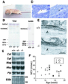
Neurological abnormalities in _Cst_−/− mice. (A) Postural abnormality of _Cst_−/− mice. The mutant mice dragged their hindlimbs during walking and their posture was flat because of the hindlimb weakness. Knockout mice were not able to grip a horizontal bar with their forelimbs, suggesting that the forelimbs are also weak. (B) Glycolipid analysis of brains from 12-week-old wild-type (+/+), heterozygous (+/−), and homozygous (−/−) littermates. Total (Left) and acidic (Right) lipid fractions, corresponding to 0.5 mg of protein, were chromatographed on precoated Silica Gel 60 HPTLC plates (Merck) by using the solvent systems: chloroform/methanol/water (60:35:8 by volume) and chloroform/methanol/0.2% CaCl2 (60:40:9 by volume), respectively, along with standard glycolipids. Orcinol/sulfuric acid was used for the detection of glycolipids. The positions of standard glycolipids are indicated on the left. (C) Northern blot analysis of myelin-marker genes. Total brain RNA from 12-week-old wild-type (+/+), heterozygous (+/−), and homozygous mutant (−/−) mice was hybridized with Cst, Cgt, Mag, Mbp, and Plp probes. Ethidium bromide (EtBr) staining of 28S and 18S ribosomal RNA is shown as a loading control. (D) Histologic analysis of cerebellum sections from a 13-week-old _Cst_−/− mouse. Myelin splitting (Left, arrow) and axonal spheroid body (Right, arrow) were observed. (E) Ultrastructure of the axoglial junction at a node of Ranvier of the myelinated fiber in cerebellar white matter from a 13-week-old wild-type (+/+) and homozygous mutant (−/−) mouse. In the mutant mouse, paranodal loops (arrows) were turned away from the axon (A). (F) Electrophysiological analysis of peripheral nerve conductivity. Maximal conduction velocity of sciatic motor nerve was measured on Cst+/+ (open square) and _Cst_−/− (closed square) mice of various ages.
Figure 3
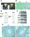
Spermatogenesis abnormalities in Cst_−/− mice. (A) Male genital organs from 8-week-old wild-type (+/+) and homozygous mutant (−/−) littermates. (B) Glycolipid analysis of testes from 8-week-old wild-type (+/+), heterozygous (+/−), and homozygous mutant (−/−) littermates. Total and acidic lipid fractions, corresponding to 2 mg of protein, were chromatographed and stained as described for Fig. 2_B. The positions of standard glycolipids are indicated on the left. (C) Histologic analysis of testis sections from 8-week-old wild-type (+/+) and mutant (−/−) littermates. (Scale bars = 50 μm.) Arrows indicate the characteristic syncytial multinucleated cells. (D) Detection of germ cell apoptosis. In situ terminal deoxynucleotidyltransferase-mediated dUTP-biotin nick end-labeling staining in the seminiferous tubules of 4-week-old wild-type (+/+) and _Cst_−/− (−/−) mice. An increase of luminal distributed apoptotic cells (brown) in the tubules of mutant testis is evident; only physiological apoptotic cells of spermatogonia are seen in wild-type testes. (Scale bars = 50 μm.) (E) RT-PCR analysis of spermatogenesis-marker genes. Total RNA from 12-week-old wild-type (+/+), heterozygous (+/−), and homozygous (−/−) testes was used as template. β-Actin RNA is shown as a loading control.
Similar articles
- Biosynthesis and biological function of sulfoglycolipids.
Honke K. Honke K. Proc Jpn Acad Ser B Phys Biol Sci. 2013;89(4):129-38. doi: 10.2183/pjab.89.129. Proc Jpn Acad Ser B Phys Biol Sci. 2013. PMID: 23574804 Free PMC article. Review. - Biological roles of sulfoglycolipids and pathophysiology of their deficiency.
Honke K, Zhang Y, Cheng X, Kotani N, Taniguchi N. Honke K, et al. Glycoconj J. 2004;21(1-2):59-62. doi: 10.1023/B:GLYC.0000043749.06556.3d. Glycoconj J. 2004. PMID: 15467400 Review. - Production of a recombinant single-chain variable-fragment (scFv) antibody against sulfoglycolipid.
Cheng X, Zhang Y, Kotani N, Watanabe T, Lee S, Wang X, Kawashima I, Tai T, Taniguchi N, Honke K. Cheng X, et al. J Biochem. 2005 Mar;137(3):415-21. doi: 10.1093/jb/mvi045. J Biochem. 2005. PMID: 15809345 - [Functions of sulfoglycolipids in membrane microdomains].
Honke K, Yamashita T. Honke K, et al. Tanpakushitsu Kakusan Koso. 2008 Sep;53(12 Suppl):1542-6. Tanpakushitsu Kakusan Koso. 2008. PMID: 21089363 Review. Japanese. No abstract available.
Cited by
- MRI characterization of paranodal junction failure and related spinal cord changes in mice.
Takano M, Hikishima K, Fujiyoshi K, Shibata S, Yasuda A, Konomi T, Hayashi A, Baba H, Honke K, Toyama Y, Okano H, Nakamura M. Takano M, et al. PLoS One. 2012;7(12):e52904. doi: 10.1371/journal.pone.0052904. Epub 2012 Dec 27. PLoS One. 2012. PMID: 23300814 Free PMC article. - Biosynthesis and biological function of sulfoglycolipids.
Honke K. Honke K. Proc Jpn Acad Ser B Phys Biol Sci. 2013;89(4):129-38. doi: 10.2183/pjab.89.129. Proc Jpn Acad Ser B Phys Biol Sci. 2013. PMID: 23574804 Free PMC article. Review. - Molecular regulators of nerve conduction - Lessons from inherited neuropathies and rodent genetic models.
Li J. Li J. Exp Neurol. 2015 May;267:209-18. doi: 10.1016/j.expneurol.2015.03.009. Epub 2015 Mar 17. Exp Neurol. 2015. PMID: 25792482 Free PMC article. Review. - A myelin galactolipid, sulfatide, is essential for maintenance of ion channels on myelinated axon but not essential for initial cluster formation.
Ishibashi T, Dupree JL, Ikenaka K, Hirahara Y, Honke K, Peles E, Popko B, Suzuki K, Nishino H, Baba H. Ishibashi T, et al. J Neurosci. 2002 Aug 1;22(15):6507-14. doi: 10.1523/JNEUROSCI.22-15-06507.2002. J Neurosci. 2002. PMID: 12151530 Free PMC article. - Myelination and regional domain differentiation of the axon.
Thaxton C, Bhat MA. Thaxton C, et al. Results Probl Cell Differ. 2009;48:1-28. doi: 10.1007/400_2009_3. Results Probl Cell Differ. 2009. PMID: 19343313 Free PMC article. Review.
References
- Humphries D E, Wong G W, Friend D S, Gurish M F, Qiu W T, Huang C, Sharpe A H, Stevens R L. Nature (London) 1999;400:769–772. - PubMed
- Forsberg E, Pejler G, Ringvall M, Lunderius C, Tomasini-Johansson B, Kusche-Gullberg M, Eriksson I, Ledin J, Hellman L, Kjellen L. Nature (London) 1999;400:773–775. - PubMed
- Akama T O, Nishida K, Nakayama J, Watanabe H, Ozaki K, Nakamura T, Dota A, Kawasaki S, Inoue Y, Maeda N, et al. Nat Genet. 2000;26:237–241. - PubMed
Publication types
MeSH terms
Substances
Grants and funding
- HD 03110/HD/NICHD NIH HHS/United States
- R01 NS024453/NS/NINDS NIH HHS/United States
- NS 27736/NS/NINDS NIH HHS/United States
- NS 24453/NS/NINDS NIH HHS/United States
- P30 HD003110/HD/NICHD NIH HHS/United States
LinkOut - more resources
Full Text Sources
Other Literature Sources
Molecular Biology Databases


