Dissecting interactions between EB1, microtubules and APC in cortical clusters at the plasma membrane - PubMed (original) (raw)
Dissecting interactions between EB1, microtubules and APC in cortical clusters at the plasma membrane
Angela I M Barth et al. J Cell Sci. 2002.
Abstract
End-binding protein (EB) 1 binds to the C-terminus of adenomatous polyposis coli (APC) protein and to the plus ends of microtubules (MT) and has been implicated in the regulation of APC accumulation in cortical clusters at the tip of extending membranes. We investigated which APC domains are involved in cluster localization and whether binding to EB1 or MTs is essential for APC cluster localization. Armadillo repeats of APC that lack EB1- and MT-binding domains are necessary and sufficient for APC localization in cortical clusters; an APC fragment lacking the armadillo repeats, but containing MT- and EB1-binding domains, does not localize to the cortical clusters but instead co-aligns with MTs throughout the cell. Significantly, analysis of endogenous proteins reveals that EB1 does not accumulate in the APC clusters. However, overexpressed EB1 does accumulate in APC clusters; the APC-binding domain in EB1 is located in the C-terminal region of EB1 between amino acids 134 and 268. Overexpressed APC- or MT-binding domains of EB1 localize to APC cortical clusters and MT, respectively, without affecting APC cluster formation itself. These results show that localization of APC in cortical clusters is different from that of EB1 at MT plus ends and appears to be independent of EB1.
Figures
Fig. 1
Subcellular distribution of endogenous APC and EB1 in MDCK cells. Basal sections of MDCK cells co-stained for EB1 (green in a–e and a″–e″) and APC (red in a′–e′ and a″–e″). Arrowheads, cortical APC clusters at the tip of cell extensions; black arrowheads, APC clusters without EB1; white arrowheads, APC clusters partially overlapping with EB1. Bars, 10 µm. Insets, higher magnification of the upper left cell extension in a-a” co-stained for EB1 (green) and APC (red). Bar, 2.5 µm.
Fig. 2
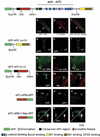
The APC armadillo repeat domain localizes to cortical APC clusters. Basal sections of cells expressing different APC domains (shown schematically on left) fused to GFP (GFP: green) co-stained for endogenous full-length APC (APC: red) or for β-tubulin (Tub: red). Note that the APC antiserum (anti-APC) does not recognize the deleted GFP-APC fusion proteins because they lack the epitope. (a–c) Full-length GFP-APC co-aligns along MTs (white arrowheads in a–c) and accumulates in cortical clusters at the tip of cell extensions distal to the MT ends (black arrowheads in a–c). (d–f) GFP-APCwoE1 co-aligns with MTs but does not colocalize with endogenous APC in clusters at the tip of cell extensions (black arrowheads in d–f). (g–n) GFP-APCE1X1 localizes to the cortical APC clusters (black arrowheads in g–i), but does not co-align with MTs (black arrowheads in k–n). (o–q) APCARM-GFP does not co-align with MTs but colocalizes with endogenous APC in the cortical clusters (black arrowheads in o–q). (r–t) APCARM-Rep.-GFP shows weak colocalization in some APC clusters (black arrowheads in r–t) but not others (white arrowheads in r–t).
Fig. 3
Binding of EB1 and the C-terminal EB1 domain to MTs or APC in vitro. (a) MT pelleting assay. Lanes 1–3 show 1/20 of the respective MBP fusion proteins after pre-clearance by centrifugation and before incubation with polymerized tubulin. Lanes 4–6 show 2/3 of the MT pellets after incubation with the respective MBP fusion proteins and centrifugation through a glycerol cushion. Lane 7, 2.5 µg purified bovine brain tubulin. (b) Binding of MBP-EB1 fusion proteins to the EB1-binding domain in GST-APCCT. Precipitation of the respective MBP fusion proteins by GST-APCCT bound to APC AB/Protein A (APC AB+GST-APCCT) (lane 7–9). GST-APCCT appears as a double band of which the faster migrating one is probably caused by partial degradation. In control assays, MBP fusion proteins were incubated with anti-GST antibody (GST AB) bound to Protein A (lane 1–3); with GST bound to GST AB/Protein A (GST AB+GST) (lane 4–6) and with anti-APC antibody (APC AB) bound to Protein A (lane 10–12). Approximately 1/15 of the total MBP fusion proteins used for each binding assay are shown in lane 13–15. M, Molecular weight standards.
Fig. 4
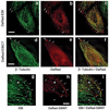
Subcellular localization of DsRed-EB1, DsRed-EB1CT and DsRedEB1NT in MDCK cells. (a–f) Basal sections of cells expressing full-length DsRed-EB1 (a–c) or DsRed-EB1CT (d–f) and co-stained with a mouse monoclonal antibody to β-tubulin (green). (a–c) DsRed-EB1 predominantly localizes to the distal (plus) ends of MTs (arrows in a–c). (d–f) DsRed-EB1CT is enriched in cortical clusters at the tip of the cell extension (arrowheads in d–f) and occasionally localizes to the plus end of MTs (arrows in d–f). (g–i) Basal sections of cell extensions of a MDCK cell expressing DsRed-EB1NT (red, marked with ‘+’) and an untransfected MDCK cell marked as ‘−’. Cells were co-stained with a mouse monoclonal antibody to EB1 (green) that does not recognize DsRed-EB1NT (see Material and Methods). DsRed-EB1NT co-aligns along MTs including their distal (plus) ends, which are marked by endogenous EB1 (green) but is not as well restricted to the distal (plus) ends of MTs as endogenous EB1 (arrows in g–i). Bar, 10 µm.
Fig. 5
Colocalization of DsRed-EB1 and DsRed-EB1CT but not of DsRed-EB1NT in cortical APC clusters. (a–i) Basal sections of cell extensions of cells expressing full-length DsRed-EB1 (a–c), DsRed-EB1NT (d–f) or DsRed-EB1CT (g–i). Cells were co-stained with APC antiserum (green). DsRed-EB1 predominantly localizes to the distal (plus) ends of MTs (arrows in b, c) (Fig. 4a–c) and colocalizes with APC in cortical clusters at the tip of the extension (white arrowheads in a–c). DsRed-EB1NT shows filamentous staining at MT distal (plus) ends and along MTs (arrows in e, f) (Fig. 4g–i) but does not colocalize with APC in cortical clusters (black arrowheads in d–f). DsRed-EB1CT colocalizes with APC at the tip of the extension (white arrowheads in g–i). Bar, 10 µm.
Similar articles
- Adenomatous polyposis coli and EB1 localize in close proximity of the mother centriole and EB1 is a functional component of centrosomes.
Louie RK, Bahmanyar S, Siemers KA, Votin V, Chang P, Stearns T, Nelson WJ, Barth AI. Louie RK, et al. J Cell Sci. 2004 Mar 1;117(Pt 7):1117-28. doi: 10.1242/jcs.00939. Epub 2004 Feb 17. J Cell Sci. 2004. PMID: 14970257 Free PMC article. - Adenomatous polyposis coli on microtubule plus ends in cell extensions can promote microtubule net growth with or without EB1.
Kita K, Wittmann T, Näthke IS, Waterman-Storer CM. Kita K, et al. Mol Biol Cell. 2006 May;17(5):2331-45. doi: 10.1091/mbc.e05-06-0498. Epub 2006 Mar 8. Mol Biol Cell. 2006. PMID: 16525027 Free PMC article. - Amer2 protein interacts with EB1 protein and adenomatous polyposis coli (APC) and controls microtubule stability and cell migration.
Pfister AS, Hadjihannas MV, Röhrig W, Schambony A, Behrens J. Pfister AS, et al. J Biol Chem. 2012 Oct 12;287(42):35333-35340. doi: 10.1074/jbc.M112.385393. Epub 2012 Aug 16. J Biol Chem. 2012. PMID: 22898821 Free PMC article. - The APC-EB1 interaction.
Morrison EE. Morrison EE. Adv Exp Med Biol. 2009;656:41-50. doi: 10.1007/978-1-4419-1145-2_4. Adv Exp Med Biol. 2009. PMID: 19928351 Review. - TIP maker and TIP marker; EB1 as a master controller of microtubule plus ends.
Vaughan KT. Vaughan KT. J Cell Biol. 2005 Oct 24;171(2):197-200. doi: 10.1083/jcb.200509150. J Cell Biol. 2005. PMID: 16247021 Free PMC article. Review.
Cited by
- Microtubule plus-ends reveal essential links between intracellular polarization and localized modulation of endocytosis during division-plane establishment in plant cells.
Dhonukshe P, Mathur J, Hülskamp M, Gadella TW Jr. Dhonukshe P, et al. BMC Biol. 2005 Apr 14;3:11. doi: 10.1186/1741-7007-3-11. BMC Biol. 2005. PMID: 15831100 Free PMC article. - The armadillo repeat domain of Apc suppresses intestinal tumorigenesis.
Crist RC, Roth JJ, Baran AA, McEntee BJ, Siracusa LD, Buchberg AM. Crist RC, et al. Mamm Genome. 2010 Oct;21(9-10):450-7. doi: 10.1007/s00335-010-9288-0. Epub 2010 Oct 1. Mamm Genome. 2010. PMID: 20886217 Free PMC article. - Coordinating cytoskeletal tracks to polarize cellular movements.
Kodama A, Lechler T, Fuchs E. Kodama A, et al. J Cell Biol. 2004 Oct 25;167(2):203-7. doi: 10.1083/jcb.200408047. J Cell Biol. 2004. PMID: 15504907 Free PMC article. Review. - Role of adenomatous polyposis coli (APC) and microtubules in directional cell migration and neuronal polarization.
Barth AI, Caro-Gonzalez HY, Nelson WJ. Barth AI, et al. Semin Cell Dev Biol. 2008 Jun;19(3):245-51. doi: 10.1016/j.semcdb.2008.02.003. Epub 2008 Feb 23. Semin Cell Dev Biol. 2008. PMID: 18387324 Free PMC article. Review. - What can humans learn from flies about adenomatous polyposis coli?
Barth AI, Nelson WJ. Barth AI, et al. Bioessays. 2002 Sep;24(9):771-4. doi: 10.1002/bies.10152. Bioessays. 2002. PMID: 12210511 Free PMC article. Review.
References
- Askham JM, Moncur P, Markham AF, Morrison EE. Regulation and function of the interaction between the APC tumour suppressor protein and EB1. Oncogene. 2000;19:1950–1958. - PubMed
- Barth AI, Nathke IS, Nelson WJ. Cadherins, catenins and APC protein: interplay between cytoskeletal complexes and signaling pathways. Curr. Opin. Cell Biol. 1997a;9:683–690. - PubMed
- Groden J, Thliveris A, Samowitz W, Carlson M, Gelbert L, Albertsen H, Joslyn G, Stevens J, Spirio L, Robertson M, et al. Identification and characterization of the familial adenomatous polyposis coli gene. Cell. 1991;66:589–600. - PubMed
Publication types
MeSH terms
Substances
LinkOut - more resources
Full Text Sources
Other Literature Sources


