Targeting assay to study the cis functions of human telomeric proteins: evidence for inhibition of telomerase by TRF1 and for activation of telomere degradation by TRF2 - PubMed (original) (raw)
Targeting assay to study the cis functions of human telomeric proteins: evidence for inhibition of telomerase by TRF1 and for activation of telomere degradation by TRF2
Katia Ancelin et al. Mol Cell Biol. 2002 May.
Abstract
We investigated the control of telomere length by the human telomeric proteins TRF1 and TRF2. To this end, we established telomerase-positive cell lines in which the targeting of these telomeric proteins to specific telomeres could be induced. We demonstrate that their targeting leads to telomere shortening. This indicates that these proteins act in cis to repress telomere elongation. Inhibition of telomerase activity by a modified oligonucleotide did not further increase the pace of telomere erosion caused by TRF1 targeting, suggesting that telomerase itself is the target of TRF1 regulation. In contrast, TRF2 targeting and telomerase inhibition have additive effects. The possibility that TRF2 can activate a telomeric degradation pathway was directly tested in human primary cells that do not express telomerase. In these cells, overexpression of full-length TRF2 leads to an increased rate of telomere shortening.
Figures
FIG. 1.
Stable introduction of LacO at chromosome ends. (A) schematic representation of the _Not_I-linearized form of the pLacOTEL plasmid. (B) Southern blot after digestion of genomic DNA with _Hin_dIII and _Msp_I and hybridization with the LacO probe. Lane 1, 182E6 cells; lane 2, LacI-expressing cells; lane 3, TRF1-LacI-expressing cells; lane 4, TRF2-LacI-expressing cells. Due to a local gel effect, the LacO bands in lanes 3 and 4 do not appear to exactly comigrate with the bands seen in lanes 1 and 2. We repeatedly noticed that these bands are identical and do not change over time (data not shown). (C) FISH of the integrated LacO sequences on a metaphase spread of the 182E6 clone with a LacO probe stained with FITC. The LacO sequence staining is indicated by open arrows. The DNA was visualized with PI in red. (D) The same metaphase shown in panel C was hybridized with probes specific for either chromosome 18 (FITC; green) or chromosome 19 (rhodamine; red) for chromosome painting. The DNA was stained with DAPI in blue. Bar = 5 μm.
FIG. 2.
Inducible expression of LacI fusion proteins. On the left, schematic structures of LacI (A), TRF1 (B), and TRF2 (C) are presented (the telobox domain is solid), as well as fusion proteins between LacI (shaded) and the nontelobox portion of TRF1 or TRF2 (hatched): TRF1-LacI or TRF2-LacI. Western analysis of inducible expression of each LacI protein in 182E6-derived cells is presented in the middle of each panel. Nuclear extracts prepared at 10 MPDs from uninduced cells (lanes 1) or clones induced with 0.1 (A and B, lanes 2) or 1 (A and B, lanes 3; C, lane 2) μM PonA were analyzed with an anti-LacI antibody (αLacI) or an anti-topoisomerase II antibody (αTopoII). Clones expressing TRF1- or TRF2-LacI under 1 μM induction (B, lane 5, and C, lane 4) or uninduced (B, lane 4, and C, lane 3) were also analyzed using an anti-TRF1 antibody (B, right) or an anti-TRF2 antibody (C, right)
FIG. 3.
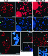
Cytological analysis of three clones derived from 182E6. Metaphase chromosomes were analyzed for LacO sequence localization. (A to C) First, in situ hybridization using a LacO FITC probe was done. (D to F) Then chromosome painting was performed, as for Fig. 1D. The DNA was visualized with PI (top panels) or DAPI (bottom panels). Metaphase chromosomes of clones expressing LacI (G), TRF1-LacI (H), and TRF2-LacI (I) were subjected to immunofluorescence analysis with an anti-LacI antibody reacting with a FITC-conjugated secondary antibody (green). The DNA was stained with PI (red). Within the insets, the same metaphase was reanalyzed using specific chromosome 18 (FITC; green) or 19 (rhodamine; red) painting probes and counterstained with DAPI. The majority of observed doublets are at, or close to, the telomeres of chromosome 18 or 19 (large arrows); strikingly, however, in TRF1-LacI and TRF2-LacI cells, but not in LacI cells, some telomeric doublets were detected that did not colocalize with either chromosome 18 or 19 (small arrows). Bars, 5 μm.
FIG. 4.
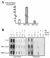
Analysis of LacI-derived protein localization by in vivo cross-linked chromatin immunoprecipitation. (A) Quantification of the immunoprecipitation of LacO DNA with anti-LacI antibody. Total and immunoprecipitated LacO DNA from cellular extracts of each LacI clone were quantified by quantitative PCR (LightCycler). Different amounts of LacO DNA associated with immunoprecipitated LacI, TRF1-LacI, or TRF2-LacI were normalized to the levels of total LacO DNA. Then, the fraction of LacO DNA associated with LacI proteins under induced conditions was compared to that under uninduced conditions. This ratio is reported on the y axis as the fold enrichment in LacO sequences. (B) Immunoprecipitation of human telomeric DNA with anti-LacI antibodies in cross-linked chromatin from 182E6-derived clones. DNA from input cellular extracts (lanes 1, 2, 1′, and 2′) and DNA immunoprecipitated (+) with anti-LacI antibodies (lanes 3, 4, 3′, and 4′) or preimmune serum (PS) (lanes 5, 6, 5′, and 6′) were blotted onto a membrane using a slot blot apparatus. The membrane was successively hybridized with either a total human genomic DNA probe (total DNA) or a TTAGGG probe. Lanes 1, 3, 5, 1′, 3′, and 5′, uninduced (0); lanes 2, 4, 6, 2′, 4′, and 6′), 1 μM PonA (1).
FIG. 5.
TRF1-LacI and TRF2-LacI effects on the lengths of the total population of telomeres. Shown are the results of TRF analysis (see Materials and Methods) of TRF1-LacI-expressing cells (A) and of TRF2-LacI-expressing cells (B). Squares, uninduced; triangles, 0.1 μM PonA; diamonds, 1 μM PonA.
FIG. 6.
cis repression of telomere elongation by TRF1-LacI or TRF2-LacI targeting. (A and D) LacI; (B and E) TRF1-LacI; (C and F) TRF2-LacI. (A to C) Examples of Southern blots. (D to F) Graphic representation of the variations of the OTRF during the experiments (rectangles, uninduced; triangles, 0.1 μM PonA; diamonds, 1 μM PonA). The dotted lines (F) correspond to values of the mean OTRF (see Materials and Methods).
FIG. 7.
OM-RNA inhibition of telomerase uncovers different mechanisms for TRF1-LacI and TRF2-LacI effects. Graphical representation of TRF and ORTF variations in TRF1-LacI (A) and TRF2-LacI (B) cells treated with 1 μM PonA during successive transfections either with no oligonucleotide (Mock experiment) or with OM-RNA (+OM-RNA). The curves correspond to the linear regressions that were used to estimate the degradation rate of telomeric DNA (the boxed values are expressed in nucleotides/MPD).
FIG. 8.
TRF2 overexpression increases the rate of telomere erosion in telomerase-negative fibroblasts. We infected WI38 cells at MPD 26 with pBloxP control or TRF2-expressing pBTRF2loxP. Genomic DNA was isolated at the indicated MPDs and analyzed for TRF length. (A) Example of _Hin_fI-_Rsa_I Southern blot used to calculate the TRF of infected WI38 cells. The asterisk indicates the location of a high-molecular-weight band (see the text). (B) Quantitative analysis of TRF during the time course of the experiment. The curves correspond to the linear regressions that were used to estimate the shortening rate of telomeric DNA (the boxed values are expressed in nucleotides per MPD).
Similar articles
- Expression of telomeric repeat binding factor 1 and 2 and TRF1-interacting nuclear protein 2 in human gastric carcinomas.
Matsutani N, Yokozaki H, Tahara E, Tahara H, Kuniyasu H, Haruma K, Chayama K, Yasui W, Tahara E. Matsutani N, et al. Int J Oncol. 2001 Sep;19(3):507-12. Int J Oncol. 2001. PMID: 11494028 - Control of human telomere length by TRF1 and TRF2.
Smogorzewska A, van Steensel B, Bianchi A, Oelmann S, Schaefer MR, Schnapp G, de Lange T. Smogorzewska A, et al. Mol Cell Biol. 2000 Mar;20(5):1659-68. doi: 10.1128/MCB.20.5.1659-1668.2000. Mol Cell Biol. 2000. PMID: 10669743 Free PMC article. - Human telomeres contain two distinct Myb-related proteins, TRF1 and TRF2.
Broccoli D, Smogorzewska A, Chong L, de Lange T. Broccoli D, et al. Nat Genet. 1997 Oct;17(2):231-5. doi: 10.1038/ng1097-231. Nat Genet. 1997. PMID: 9326950 - Regulation of telomerase by telomeric proteins.
Smogorzewska A, de Lange T. Smogorzewska A, et al. Annu Rev Biochem. 2004;73:177-208. doi: 10.1146/annurev.biochem.73.071403.160049. Annu Rev Biochem. 2004. PMID: 15189140 Review. - Post-translational modifications of TRF1 and TRF2 and their roles in telomere maintenance.
Walker JR, Zhu XD. Walker JR, et al. Mech Ageing Dev. 2012 Jun;133(6):421-34. doi: 10.1016/j.mad.2012.05.002. Epub 2012 May 23. Mech Ageing Dev. 2012. PMID: 22634377 Review.
Cited by
- Telomere length modulation in human astroglial brain tumors.
La Torre D, Conti A, Aguennouz MH, De Pasquale MG, Romeo S, Angileri FF, Cardali S, Tomasello C, Alafaci C, Germanò A. La Torre D, et al. PLoS One. 2013 May 14;8(5):e64296. doi: 10.1371/journal.pone.0064296. Print 2013. PLoS One. 2013. PMID: 23691191 Free PMC article. - Human telomerase and its regulation.
Cong YS, Wright WE, Shay JW. Cong YS, et al. Microbiol Mol Biol Rev. 2002 Sep;66(3):407-25, table of contents. doi: 10.1128/MMBR.66.3.407-425.2002. Microbiol Mol Biol Rev. 2002. PMID: 12208997 Free PMC article. Review. - Telomere rapid deletion regulates telomere length in Arabidopsis thaliana.
Watson JM, Shippen DE. Watson JM, et al. Mol Cell Biol. 2007 Mar;27(5):1706-15. doi: 10.1128/MCB.02059-06. Epub 2006 Dec 22. Mol Cell Biol. 2007. PMID: 17189431 Free PMC article. - Dynamics of protein binding to telomeres in living cells: implications for telomere structure and function.
Mattern KA, Swiggers SJ, Nigg AL, Löwenberg B, Houtsmuller AB, Zijlmans JM. Mattern KA, et al. Mol Cell Biol. 2004 Jun;24(12):5587-94. doi: 10.1128/MCB.24.12.5587-5594.2004. Mol Cell Biol. 2004. PMID: 15169917 Free PMC article. - NuMA is a major acceptor of poly(ADP-ribosyl)ation by tankyrase 1 in mitosis.
Chang W, Dynek JN, Smith S. Chang W, et al. Biochem J. 2005 Oct 15;391(Pt 2):177-84. doi: 10.1042/BJ20050885. Biochem J. 2005. PMID: 16076287 Free PMC article.
References
- Artandi, S. E., and R. A. DePinho. 2000. A critical role for telomeres in suppressing and facilitating carcinogenesis. Curr. Opin. Genet. Dev. 10:39-46. - PubMed
- Baumann, P., and T. R. Cech. 2001. Pot1, the putative telomere end-binding protein in fission yeast and humans. Science 292:1171-1175. - PubMed
- Beattie, T. L., W. Zhou, M. O. Robinson, and L. Harrington. 1998. Reconstitution of human telomerase activity in vitro. Curr. Biol. 8:177-180. - PubMed
- Bilaud, T., C. Brun, K. Ancelin, C. E. Koering, T. Laroche, and E. Gilson. 1997. Telomeric localization of TRF2, a novel human telobox protein. Nat. Genet. 17:236-239. - PubMed
Publication types
MeSH terms
Substances
LinkOut - more resources
Full Text Sources
Other Literature Sources
Molecular Biology Databases
Research Materials
Miscellaneous


