Diffusion tensor MR imaging in diffuse axonal injury - PubMed (original) (raw)
. 2002 May;23(5):794-802.
Affiliations
- PMID: 12006280
- PMCID: PMC7974716
Diffusion tensor MR imaging in diffuse axonal injury
Konstantinos Arfanakis et al. AJNR Am J Neuroradiol. 2002 May.
Abstract
Background and purpose: Disruption of the cytoskeletal network and axonal membranes characterizes diffuse axonal injury (DAI) in the first few hours after traumatic brain injury. Histologic abnormalities seen in DAI hypothetically decrease the diffusion along axons and increase the diffusion in directions perpendicular to them. DAI therefore is hypothetically associated in the short term with decreased diffusion anisotropy. We tested this hypothesis by measuring the diffusion characteristics of traumatized brain tissue with use of diffusion tensor MR imaging.
Methods: Five patients with mild traumatic brain injuries and 10 control subjects were studied with CT, conventional MR imaging, and diffusion tensor imaging. All patients were examined within 24 hours of injury. In each participant, diffusion tensor indices from homologous normal-appearing white matter regions of both hemispheres were compared. These indices were also compared between homologous regions of each patient and the control group. In two patients, diffusion tensor images from the immediate post-trauma period were compared with those at 1 month follow-up.
Results: Patients displayed significant reduction of diffusion anisotropy in several regions compared with the homologous ones in the contralateral hemisphere. Such differences were not observed in the control subjects. Significant reduction of diffusion anisotropy was also detected when diffusion tensor results from the patients were compared with those of the controls. This reduction was often less evident 1 month after injury.
Conclusion: White matter regions with reduced anisotropy are detected in the first 24 hours after traumatic brain injury. Therefore, diffusion tensor imaging may be a powerful technique for in vivo detection of DAI.
Figures
Fig 1.
Illustration of the changes that axons undergo owing to cytoskeletal perturbation from mild traumatic brain injury. A, The top neuron is healthy. In the bottom neuron, neurofilamentous and, generally, cytoskeletal misalignment is visible a short time after injury. This impairs axonal transport. B, Organelles accumulate in the injured region, causing the axon to swell locally and subsequently disconnect from the rest. In this Figure, the dimensions of the axons relative to the interaxonal space do not necessarily correspond to reality.
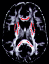
Fig 2.
Example of the selection of voxels from the five structures under study, in one section of a control subject. The background image is an LI map. The red dots correspond to the selected voxels. The dots were made larger than the in-plane dimensions of a single voxel for visualization purposes.
Fig 3.
A, LI map in a control subject. The border between the anterior (AIC) and posterior (PIC) limbs of the internal capsule is not obvious from this image. B, Absolute value color map of the same section as in A. Arrows indicate the exact level of the border between AIC and PIC. Color representation of directions in 3D space helps to clearly separate the two structures. C, Color circle demonstrates the correspondence of colors and directions in 3D space. This circle should be thought of as a 3D dome that is viewed from below.
Fig 4.
A, T2-weighted image in a patient with mild traumatic brain injury. Small hemorrhagic lesions are visible in the left occipital lobe and right frontal lobe. B, Diffusion-weighted map of the same section in the same patient, for one of the 23 diffusion-weighted gradient directions. The abnormal regions from A are also visible in B. C, Corresponding trace map. The same lesions from A and B can be detected. D, LI map of the same section in the same patient. Decreased anisotropy is visible in the left internal capsule and left anterior corpus callosum. No abnormality was demonstrated in these regions with any other type of imaging. On this LI map, however, the hemorrhagic lesions from A, B, and C are hard to identify. R indicates right; L, left.
Fig 5.
Graphs show distributions of FA results from 10 selected ROIs (5 structures x 2 hemispheres) for the control subjects (continuous lines) combined with the corresponding FA values for the five patients with mild traumatic brain injuries (vertical lines), in the left (A) and right (B) hemispheres. All horizontal axes represent FA values. P values are reported only for cases in which FA results for the patients were significantly different (P < .05) from the mean FA results of the control subjects. For the patients, anisotropy was found to be significantly reduced in several structures compared with normal values. Reduction of FA was more often in the internal capsule and corpus callosum and less often in the external capsule of the patients. LACC and RACC indicate left and right anterior corpus callosum, respectively; LPCC and RPCC, left and right posterior corpus callosum; LEC and REC, left and right external capsule; LAIC and RAIC, left and right anterior internal capsule; LPIC and RPIC, left and right posterior internal capsule.
Fig 6.
Bar graphs of ratios of diffusion characteristics between the two sides of four structures in a patient with mild traumatic brain injury, using the immediate (A) and short-term follow-up (B) data. The quantities λ1, λ2, λ3, trace, FA, and LI correspond to that side of the structure that has the lowest anisotropy on the immediate image. The quantities λ1’, λ2’, λ3’, trace’, FA’, and LI’ correspond to the contralateral side of these structures. The four structures are external capsule (EC), anterior corpus callosum (ACC), posterior internal capsule (PIC), and anterior internal capsule (AIC). Each structure corresponds to a specific color. For the immediate data set, one side of the four structures has lower λ1, higher λ2 and λ3, and lower FA and LI than its contralateral homologous side. Also, trace is similar for both sides. For the follow-up data set, λ1, λ2, λ3, FA, and LI have become more similar between the two sides of each structure, therefore giving ratios that are closer to 1. The ratios of the trace have remained at values almost equal to 1. When looking at the absolute values of the diffusion characteristics, we notice that the ratios change because the side of each structure that had low anisotropy on the immediate dataset has higher anisotropy on the follow-up dataset.
Similar articles
- Diffuse axonal injury has a characteristic multidimensional MRI signature in the human brain.
Benjamini D, Iacono D, Komlosh ME, Perl DP, Brody DL, Basser PJ. Benjamini D, et al. Brain. 2021 Apr 12;144(3):800-816. doi: 10.1093/brain/awaa447. Brain. 2021. PMID: 33739417 Free PMC article. - Diffuse axonal injury in severe traumatic brain injury visualized using high-resolution diffusion tensor imaging.
Xu J, Rasmussen IA, Lagopoulos J, Håberg A. Xu J, et al. J Neurotrauma. 2007 May;24(5):753-65. doi: 10.1089/neu.2006.0208. J Neurotrauma. 2007. PMID: 17518531 - Quantitative assessments of traumatic axonal injury in human brain: concordance of microdialysis and advanced MRI.
Magnoni S, Mac Donald CL, Esparza TJ, Conte V, Sorrell J, Macrì M, Bertani G, Biffi R, Costa A, Sammons B, Snyder AZ, Shimony JS, Triulzi F, Stocchetti N, Brody DL. Magnoni S, et al. Brain. 2015 Aug;138(Pt 8):2263-77. doi: 10.1093/brain/awv152. Epub 2015 Jun 17. Brain. 2015. PMID: 26084657 Free PMC article. - [Application of diffusion tensor imaging and 1H-magnetic resonance spectroscopy in diagnosis of traumatic brain injury].
Zhao Z, Yu JY, Wu KH, Yu HL, Liu AX, Li YH. Zhao Z, et al. Fa Yi Xue Za Zhi. 2012 Jun;28(3):207-10. Fa Yi Xue Za Zhi. 2012. PMID: 22812225 Review. Chinese. - Current contribution of diffusion tensor imaging in the evaluation of diffuse axonal injury.
Grassi DC, Conceição DMD, Leite CDC, Andrade CS. Grassi DC, et al. Arq Neuropsiquiatr. 2018 Mar;76(3):189-199. doi: 10.1590/0004-282x20180007. Arq Neuropsiquiatr. 2018. PMID: 29809237 Review.
Cited by
- A decade of DTI in traumatic brain injury: 10 years and 100 articles later.
Hulkower MB, Poliak DB, Rosenbaum SB, Zimmerman ME, Lipton ML. Hulkower MB, et al. AJNR Am J Neuroradiol. 2013 Nov-Dec;34(11):2064-74. doi: 10.3174/ajnr.A3395. Epub 2013 Jan 10. AJNR Am J Neuroradiol. 2013. PMID: 23306011 Free PMC article. Review. - Altered DTI scalars in the hippocampus are associated with morphological and structural changes after traumatic brain injury.
Arora P, Trivedi R, Kumari M, Singh K, Sandhir R, D'Souza MM, Rana P. Arora P, et al. Brain Struct Funct. 2024 May;229(4):853-863. doi: 10.1007/s00429-024-02758-8. Epub 2024 Feb 21. Brain Struct Funct. 2024. PMID: 38381381 - Hyper-connectivity of the thalamus during early stages following mild traumatic brain injury.
Sours C, George EO, Zhuo J, Roys S, Gullapalli RP. Sours C, et al. Brain Imaging Behav. 2015 Sep;9(3):550-63. doi: 10.1007/s11682-015-9424-2. Brain Imaging Behav. 2015. PMID: 26153468 Free PMC article. - MR Imaging Applications in Mild Traumatic Brain Injury: An Imaging Update.
Wu X, Kirov II, Gonen O, Ge Y, Grossman RI, Lui YW. Wu X, et al. Radiology. 2016 Jun;279(3):693-707. doi: 10.1148/radiol.16142535. Radiology. 2016. PMID: 27183405 Free PMC article. - A review of magnetic resonance imaging and diffusion tensor imaging findings in mild traumatic brain injury.
Shenton ME, Hamoda HM, Schneiderman JS, Bouix S, Pasternak O, Rathi Y, Vu MA, Purohit MP, Helmer K, Koerte I, Lin AP, Westin CF, Kikinis R, Kubicki M, Stern RA, Zafonte R. Shenton ME, et al. Brain Imaging Behav. 2012 Jun;6(2):137-92. doi: 10.1007/s11682-012-9156-5. Brain Imaging Behav. 2012. PMID: 22438191 Free PMC article. Review.
References
- Kraus J, Nourjah P. The epidemiology of mild head injury. In: Levin H, Eisenberg H, Benton A, eds. Mild Head Injury. Oxford: Oxford University Press;1989. :8–22
- Capruso DX, Levin HS. Cognitive impairment following closed head injury. Neurol Clin 1992;10:879–893 - PubMed
- Evans RW. The postconcussion syndrome and the sequelae of mild head injury. Neurol Clin 1992;10:815–847 - PubMed
- Levin HS, Mattis S, Ruff RM, et al. Neurobehavioral outcome following minor head injury: a three-center study. J Neurosurg 1987;66:234–243 - PubMed
MeSH terms
LinkOut - more resources
Full Text Sources
Medical


