The effect of Replens on vaginal cytology in the treatment of postmenopausal atrophy: cytomorphology versus computerised cytometry - PubMed (original) (raw)
Clinical Trial
The effect of Replens on vaginal cytology in the treatment of postmenopausal atrophy: cytomorphology versus computerised cytometry
J A W M van der Laak et al. J Clin Pathol. 2002 Jun.
Abstract
Background: After the menopause decreased concentrations of oestrogen may result in insufficient maturation of the vaginal epithelium, which can lead to a range of vaginal discomforts. This state of vaginal atrophy may be treated with oestrogen replacement treatment. Replens, a non-hormonal alternative to oestrogen replacement treatment has been shown to be effective in relieving symptoms related to vaginal atrophy in previous studies.
Aims: To study the effect of Replens on the maturation of the vaginal epithelium and morphology of the vaginal cells and to compare the results of a recently developed cytomorphometric method with manual assessment of the degree of maturation in vaginal smears.
Methods: Vaginal smears from 38 postmenopausal women suffering from symptoms related to vaginal atrophy were analysed manually and by cytomorphometry. The maturation value (MV) and the percentages of (para)basal, intermediate, and superficial cells (maturation index; MI) were measured by both methods before and after treatment with Replens. Cytomorphometry also measured mean cellular area, mean nuclear area, and mean area ratio.
Results: A correlation was shown between the two methods in the assessment of percentages of (para) basal and intermediate cells and MV. Cytomorphometric data showed a significant increase in mean cellular area, indicating a positive effect of Replens on the maturation of the vaginal epithelium. Changes in nuclear area and ratio between nuclear and cellular areas were not significant. Treatment with Replens did not influence MI or MV, as assessed by the two methods.
Conclusions: Replens did have an effect on vaginal morphology. The automated procedure may be useful for the assessment of maturation in vaginal smears and is more sensitive to small (subvisual) changes.
Figures
Figure 1
Comparison between automated and manual assessment of baseline values for (A) the percentage of (para)basal cells and (B) the maturation value.
Figure 2
Difference between post treatment and baseline values for (A) the percentage of (para)basal cells, (B) the maturation value, and (C) the cellular area. The solid lines indicate the median values over all cases.
Figure 3
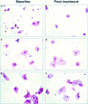
Randomly selected microscopical images of haematoxylin and eosin stained smears from three women with a pronounced increase in cellular area, as assessed by cytomorphometric analysis. Patients 1 (A, B), 2 (C, D), and 3 (E, F) at baseline (A, C, E) and post treatment (B, D, F).
Similar articles
- Replens versus dienoestrol cream in the symptomatic treatment of vaginal atrophy in postmenopausal women.
Bygdeman M, Swahn ML. Bygdeman M, et al. Maturitas. 1996 Apr;23(3):259-63. doi: 10.1016/0378-5122(95)00955-8. Maturitas. 1996. PMID: 8794418 Clinical Trial. - The effect of oxytocin vaginal gel on vaginal atrophy in postmenopausal women: a randomized controlled trial.
Zohrabi I, Abedi P, Ansari S, Maraghi E, Shakiba Maram N, Houshmand G. Zohrabi I, et al. BMC Womens Health. 2020 May 19;20(1):108. doi: 10.1186/s12905-020-00935-5. BMC Womens Health. 2020. PMID: 32429977 Free PMC article. Clinical Trial. - Comparative study: Replens versus local estrogen in menopausal women.
Nachtigall LE. Nachtigall LE. Fertil Steril. 1994 Jan;61(1):178-80. doi: 10.1016/s0015-0282(16)56474-7. Fertil Steril. 1994. PMID: 8293835 Clinical Trial. - The role of local vaginal estrogen for treatment of vaginal atrophy in postmenopausal women: 2007 position statement of The North American Menopause Society.
North American Menopause Society. North American Menopause Society. Menopause. 2007 May-Jun;14(3 Pt 1):355-69; quiz 370-1. doi: 10.1097/gme.0b013e31805170eb. Menopause. 2007. PMID: 17438512 - An overview of the phytoestrogen effect on vaginal health and dyspareunia in peri- and post-menopausal women.
Dizavandi FR, Ghazanfarpour M, Roozbeh N, Kargarfard L, Khadivzadeh T, Dashti S. Dizavandi FR, et al. Post Reprod Health. 2019 Mar;25(1):11-20. doi: 10.1177/2053369118823365. Epub 2019 Feb 20. Post Reprod Health. 2019. PMID: 30786797 Review.
Cited by
- The effect of vitamin D on vaginal atrophy in postmenopausal women.
Rad P, Tadayon M, Abbaspour M, Latifi SM, Rashidi I, Delaviz H. Rad P, et al. Iran J Nurs Midwifery Res. 2015 Mar-Apr;20(2):211-5. Iran J Nurs Midwifery Res. 2015. PMID: 25878698 Free PMC article. - Impact of vulvovaginal health on postmenopausal women: a review of surveys on symptoms of vulvovaginal atrophy.
Parish SJ, Nappi RE, Krychman ML, Kellogg-Spadt S, Simon JA, Goldstein JA, Kingsberg SA. Parish SJ, et al. Int J Womens Health. 2013 Jul 29;5:437-47. doi: 10.2147/IJWH.S44579. Print 2013. Int J Womens Health. 2013. PMID: 23935388 Free PMC article. - Study of Vulvovaginal Atrophy and Genitourinary Syndrome of Menopause and Its Impact on the Quality of Life of Postmenopausal Women in Central India.
Ulhe SC, Acharya N, Vats A, Singh A. Ulhe SC, et al. Cureus. 2024 Feb 24;16(2):e54802. doi: 10.7759/cureus.54802. eCollection 2024 Feb. Cureus. 2024. PMID: 38529421 Free PMC article. - Maintaining sexual health throughout gynecologic cancer survivorship: A comprehensive review and clinical guide.
Huffman LB, Hartenbach EM, Carter J, Rash JK, Kushner DM. Huffman LB, et al. Gynecol Oncol. 2016 Feb;140(2):359-68. doi: 10.1016/j.ygyno.2015.11.010. Epub 2015 Nov 7. Gynecol Oncol. 2016. PMID: 26556768 Free PMC article. Review. - Topical testosterone for breast cancer patients with vaginal atrophy related to aromatase inhibitors: a phase I/II study.
Witherby S, Johnson J, Demers L, Mount S, Littenberg B, Maclean CD, Wood M, Muss H. Witherby S, et al. Oncologist. 2011;16(4):424-31. doi: 10.1634/theoncologist.2010-0435. Epub 2011 Mar 8. Oncologist. 2011. PMID: 21385795 Free PMC article. Clinical Trial.
References
- Kaufman RH, Friedrich EG, Gardner HL. Atrophic, desquamative, and postradiation vulvovaginitis. In: Benign diseases of the vulva and vagina. Chicago: Year book medical publishers, 1989:419–24.
- Nachtigall LE. Comparative study: Replens versus local estrogen in menopausal women. Fertil Steril 1994;61:178–80. - PubMed
- Bygdeman M, Swahn ML. Replens versus dienoestrol cream in the symptomatic treatment of vaginal atrophy in postmenopausal women. Maturitas 1996;23:259–63. - PubMed
- Hubbard GB, Carey KD Levine H, et al. Evaluation of a vaginal moisturizer in baboons with decreasing ovarian function. Lab Anim Sci 1997;47:36–9. - PubMed
- Gelfand MM, Wendman E. Treating vaginal dryness in breast cancer patients: results of applying a polycarbophil moisturizing gel. J Womens Health 1994;3:427–34.
Publication types
MeSH terms
Substances
LinkOut - more resources
Full Text Sources
Other Literature Sources


