Prefrontal cortical volume reduction associated with frontal cortex function deficit in 6-week abstinent crack-cocaine dependent men - PubMed (original) (raw)
Comparative Study
Prefrontal cortical volume reduction associated with frontal cortex function deficit in 6-week abstinent crack-cocaine dependent men
George Fein et al. Drug Alcohol Depend. 2002.
Abstract
Background: This study examined regional cortical volumes in 6-week abstinent men dependent on crack-cocaine only (Cr) or on both crack-cocaine and alcohol (CrA). Our goal was to test the a priori hypothesis of prefrontal cortical volume reduction, along with associated impairments in frontal mediated functions, and to look for differences between the Cr and CrA groups.
Methods: Structural magnetic resonance imaging (MRI) of the brain and neuropsychological assessment were performed on 17 6-week abstinent Cr subjects, 29 six-week abstinent CrA subjects, and 20 normal controls. Cortical volume was quantified in the prefrontal, parietal, temporal and occipital regions.
Results: Cr and CrA subjects showed comparable reductions in prefrontal gray matter volume compared to controls; this reduction was negatively associated with performance impairments in the executive function domain.
Conclusions: Dependence on Cr (with or without concomitant alcohol dependence) was associated with reduced prefrontal cortical volume. Cr dependence with concomitant alcohol dependence was not associated with greater prefrontal volume reductions than Cr dependence alone. The existence of these findings at 6-week abstinence indicates that they are not a result of acute cocaine or alcohol exposure. The association of reduced prefrontal cortical volume with cognitive impairments in frontal cortex mediated abilities suggests that this reduced cerebral volume has functional consequences.
Figures
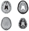
Fig. 1
Counter-clockwise from the top right: the same mid-ventricular axial MRI slice in the T2-weighted, T1-weighted, PD-weighted, and segmented forms for an abstinent Cr and alcohol dependent subject (age 52). The segmented image shows a moderate amount of white matter signal hyperintensity (white areas).
Fig. 2
The abstinent substance dependent samples had reduced regional gray matter in the prefrontal cortex only, with group membership accounting for 10.0% of the variance (P <_ 0.01); this is compared to less than 1.6% of the variance for parietal, temporal, and occipital cortices (all _P > 0.32).
Similar articles
- Association of frontal and posterior cortical gray matter volume with time to alcohol relapse: a prospective study.
Rando K, Hong KI, Bhagwagar Z, Li CS, Bergquist K, Guarnaccia J, Sinha R. Rando K, et al. Am J Psychiatry. 2011 Feb;168(2):183-92. doi: 10.1176/appi.ajp.2010.10020233. Epub 2010 Nov 15. Am J Psychiatry. 2011. PMID: 21078704 Free PMC article. - Neuropsychological performance of individuals dependent on crack-cocaine, or crack-cocaine and alcohol, at 6 weeks and 6 months of abstinence.
Di Sclafani V, Tolou-Shams M, Price LJ, Fein G. Di Sclafani V, et al. Drug Alcohol Depend. 2002 Apr 1;66(2):161-71. doi: 10.1016/s0376-8716(01)00197-1. Drug Alcohol Depend. 2002. PMID: 11906803 Free PMC article. - [Structural and functional neuroanatomy of attention-deficit hyperactivity disorder (ADHD)].
Emond V, Joyal C, Poissant H. Emond V, et al. Encephale. 2009 Apr;35(2):107-14. doi: 10.1016/j.encep.2008.01.005. Epub 2008 Jul 7. Encephale. 2009. PMID: 19393378 Review. French.
Cited by
- Smaller regional gray matter volume in homeless african american cocaine-dependent men: a preliminary report.
Weller RE, Stoeckel LE, Milby JB, Bolding M, Twieg DB, Knowlton RC, Avison MJ, Ding Z. Weller RE, et al. Open Neuroimag J. 2011;5:57-64. doi: 10.2174/1874440001105010057. Epub 2011 Oct 28. Open Neuroimag J. 2011. PMID: 22135719 Free PMC article. - Lower cortical thickness and increased brain aging in adults with cocaine use disorder.
Schinz D, Schmitz-Koep B, Tahedl M, Teckenberg T, Schultz V, Schulz J, Zimmer C, Sorg C, Gaser C, Hedderich DM. Schinz D, et al. Front Psychiatry. 2023 Nov 13;14:1266770. doi: 10.3389/fpsyt.2023.1266770. eCollection 2023. Front Psychiatry. 2023. PMID: 38025412 Free PMC article. - Neurocognition and inhibitory control in polysubstance use disorders: Comparison with alcohol use disorders and changes with abstinence.
Schmidt TP, Pennington DL, Cardoos SL, Durazzo TC, Meyerhoff DJ. Schmidt TP, et al. J Clin Exp Neuropsychol. 2017 Feb;39(1):22-34. doi: 10.1080/13803395.2016.1196165. Epub 2016 Oct 3. J Clin Exp Neuropsychol. 2017. PMID: 27690739 Free PMC article. - Prior chronic cocaine exposure in mice induces persistent alterations in cognitive function.
Krueger DD, Howell JL, Oo H, Olausson P, Taylor JR, Nairn AC. Krueger DD, et al. Behav Pharmacol. 2009 Dec;20(8):695-704. doi: 10.1097/FBP.0b013e328333a2bb. Behav Pharmacol. 2009. PMID: 19901826 Free PMC article. - Reduced frontal brain volume in non-treatment-seeking cocaine-dependent individuals: exploring the role of impulsivity, depression, and smoking.
Crunelle CL, Kaag AM, van Wingen G, van den Munkhof HE, Homberg JR, Reneman L, van den Brink W. Crunelle CL, et al. Front Hum Neurosci. 2014 Jan 17;8:7. doi: 10.3389/fnhum.2014.00007. eCollection 2014. Front Hum Neurosci. 2014. PMID: 24478673 Free PMC article.
References
- Bartzokis G, Beckson M, Lu P, Edwards N, Rapoport R, Wiseman E, Bridge P. Age-related brain volume reductions in amphetamine and cocaine addicts and normal controls: implications for addiction research. Psychiat Res: Neuroimag. 2000;98:93–102. - PubMed
- Berman S, Noble E. Reduced visuospatial performance in children with the D2 dopamine receptor A1 allele. Behav Genet. 1995;25 (1):45–58. - PubMed
- Biggins C, Fein G, MacKay S. ERP evidence for frontal cortex effects of chronic cocaine abuse. Biol Psychiat. 1997;42 (6):472–485. - PubMed
- Brown TG, Seraganian P, Tremblay J. Alcoholics also dependent on cocaine in treatment: do they differ from ‘pure’ alcoholics? Addict Behav. 1994;19 (1):105–112. - PubMed
Publication types
MeSH terms
Substances
LinkOut - more resources
Full Text Sources
Medical

