Differing mechanisms for glutamate receptor aggregation on dendritic spines and shafts in cultured hippocampal neurons - PubMed (original) (raw)
Differing mechanisms for glutamate receptor aggregation on dendritic spines and shafts in cultured hippocampal neurons
Ruifa Mi et al. J Neurosci. 2002.
Abstract
We have explored the ability of axons from spinal and hippocampal neurons to aggregate NMDA- and AMPA-type glutamate receptors on each other as a way of exploring the molecular differences between their presynaptic elements. Spinal axons, which normally cluster only AMPA-type glutamate receptors on other spinal neurons, cluster both AMPA- and NMDA-type glutamate receptors on the dendritic shafts of hippocampal interneurons but are ineffective at clustering either subtype of glutamate receptor on the dendritic spines of hippocampal pyramidal neurons. Conversely, hippocampal axons appear to be multipotent, capable of clustering both AMPA- and NMDA-type glutamate receptors on hippocampal interneurons and pyramidal cells. The secretion of the neuronal activity-regulated pentraxin (Narp) by hippocampal axons is restricted to contacts with interneurons. Exogenous application of Narp to cultured hippocampal neurons results in clusters of both NMDA- and AMPA-type glutamate receptors on hippocampal interneurons but not hippocampal pyramidal neurons. Because Narp displays no ability to directly aggregate NMDA receptors, we propose that Narp aggregates NMDA receptors in hippocampal interneurons indirectly through cytoplasmic coupling to synaptic AMPA receptors. Furthermore, our data suggest the existence of a novel molecule(s), capable of forming excitatory synapses on dendritic spines.
Figures
Fig. 1.
The distribution of glutamate receptors in cultured hippocampal and spinal neurons. Neurons grown for 2 weeks_in vitro_ were fixed and permeabilized as described in Materials and Methods and stained with antibodies to the glutamate receptor subunits GluR1 and NR1 and the presynaptic vesicle protein synaptophysin (Syn). Although NR1 and GluR1 showed close colocalization on the spines (A, B) and shafts (E, F) of hippocampal neurons, there was no colocalization in spinal neurons (I, J). Nearly all clusters of NR1 or GluR1 were associated with synaptophysin. Scale bars, 5 μm.
Fig. 2.
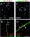
Differing capabilities of spinal and hippocampal axons to cluster glutamate receptors. A,B, A transfected (GFP positive) hippocampal axon (Hip) contacts a spiny hippocampal pyramidal cell stained with GluR2 (red). Two GluR2-positive spines (arrows) are seen to colocalize with the transfected axon. C, D, A transfected spinal cord axon contacts another spiny hippocampal pyramidal neuron. In this case no spine or shaft (arrowheads) clusters of GluR2 are seen to colocalize with the axon. Scale bar, 5 μm.
Fig. 3.
NMDA receptor aggregation is determined by the postsynaptic cell. A, B, A GFP-positive spinal axon contacts a hippocampal interneuron and is associated with a cluster of NR1. C, D, A GFP-positive hippocampal axon contacts a spinal neuron and is associated with several clusters of GluR2 (arrows). E–G, Another hippocampal axon contacts a spinal neuron and is not associated with clustered NR1. The spinal neuron is positively identified by the nuclear EYFP staining. H, I, Total and surface (biotinylated) NR1, GluR1, and GluR2 (total only) are shown from day 14 in vitro cultures of hippocampal and spinal neurons, demonstrating that the surface NR1 expression in spinal (SC) and hippocampal cultures (HIP) is nearly equivalent, especially by comparison with GluR1 and GluR2.
Fig. 4.
Hippocampal axons localize mycNarp exclusively at interneuron synapses. A–C, A hippocampal axon from a neuron transfected with GFP and mycNarp contacts an interneuron. A cluster of surface mycNarp (green) is seen to colocalize with a GluR2 cluster (red) (C). D–F, Another transfected hippocampal axon contacts a spiny pyramidal cell where it colocalizes with two GluR2 clusters (D, E,arrows). Two small clusters of mycNarp (green) appear along the axon (F) but do not colocalize with the clustered spiny GluR2 staining.
Fig. 5.
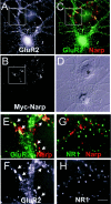
Narp clusters GluR1 but not NR1 on hippocampal pyramidal neurons. A–F, A mycNarp-transfected 293 cell (B, asterisk in D) contacts a GluR1-positive pyramidal neuron (A).C, Sites of overlap between mycNarp and GluR1 are seen (yellow). Magnified views of the boxed areas indicated in A and C are shown in F and E, respectively. The colocalizing clusters of mycNarp and GluR1 are indicated by_arrows_. G, H, Another 293 cell transfected with mycNarp showed no colocalization of NR1 immmunostaining (green) with mycNarp (red).
Fig. 6.
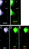
Narp clusters NR1 and GluR2 on hippocampal interneurons. A, B, Surface mycNarp from a transfected 293 cell (red) is seen to colocalize with two clusters of GluR2 (green) on a contacted interneuron (arrows). C–E, Another interneuron, this time stained with an antibody to NR1, is contacted by another mycNarp-secreting 293 cell. Multiple sites of mycNarp (red) and NR1 (green) overlap are seen (C, D,asterisk). In E, the sites of mycNarp–NR1 overlap are devoid of presynaptic synaptophysin immunostaining (red).
Fig. 7.
Narp does not cluster NR1 in spinal neurons or HEK 293 cells. A, B, The dendrite of a spinal neuron immunostained with an antibody to NR1 is contacted by a mycNarp-expressing 293 cell (B,red). No coclustering of NR1 is seen.C–F, A 293 cell expressing Narp (red), HA-tagged GluR1 (blue), NR2A (untagged), and myc-tagged NR1 (green) is seen to colocalize GluR1 and Narp (C, D) but not NR1 and Narp (F).
Similar articles
- Synaptically targeted narp plays an essential role in the aggregation of AMPA receptors at excitatory synapses in cultured spinal neurons.
O'Brien R, Xu D, Mi R, Tang X, Hopf C, Worley P. O'Brien R, et al. J Neurosci. 2002 Jun 1;22(11):4487-98. doi: 10.1523/JNEUROSCI.22-11-04487.2002. J Neurosci. 2002. PMID: 12040056 Free PMC article. - Mismatched appositions of presynaptic and postsynaptic components in isolated hippocampal neurons.
Rao A, Cha EM, Craig AM. Rao A, et al. J Neurosci. 2000 Nov 15;20(22):8344-53. doi: 10.1523/JNEUROSCI.20-22-08344.2000. J Neurosci. 2000. PMID: 11069941 Free PMC article. - PSD-95 involvement in maturation of excitatory synapses.
El-Husseini AE, Schnell E, Chetkovich DM, Nicoll RA, Bredt DS. El-Husseini AE, et al. Science. 2000 Nov 17;290(5495):1364-8. Science. 2000. PMID: 11082065 - Glutamate receptor dynamics in dendritic microdomains.
Newpher TM, Ehlers MD. Newpher TM, et al. Neuron. 2008 May 22;58(4):472-97. doi: 10.1016/j.neuron.2008.04.030. Neuron. 2008. PMID: 18498731 Free PMC article. Review. - Hippocampal neurons in schizophrenia.
Heckers S, Konradi C. Heckers S, et al. J Neural Transm (Vienna). 2002 May;109(5-6):891-905. doi: 10.1007/s007020200073. J Neural Transm (Vienna). 2002. PMID: 12111476 Free PMC article. Review.
Cited by
- Activity-dependent movements of postsynaptic scaffolds at inhibitory synapses.
Hanus C, Ehrensperger MV, Triller A. Hanus C, et al. J Neurosci. 2006 Apr 26;26(17):4586-95. doi: 10.1523/JNEUROSCI.5123-05.2006. J Neurosci. 2006. PMID: 16641238 Free PMC article. - Transsynaptic channelosomes: non-conducting roles of ion channels in synapse formation.
Nishimune H. Nishimune H. Channels (Austin). 2011 Sep-Oct;5(5):432-9. doi: 10.4161/chan.5.5.16472. Epub 2011 Sep 1. Channels (Austin). 2011. PMID: 21654201 Free PMC article. Review. - Histone Deacetylase 2 Inhibition Attenuates Downregulation of Hippocampal Plasticity Gene Expression during Aging.
Singh P, Thakur MK. Singh P, et al. Mol Neurobiol. 2018 Mar;55(3):2432-2442. doi: 10.1007/s12035-017-0490-x. Epub 2017 Mar 31. Mol Neurobiol. 2018. PMID: 28364391 - Organization of amyloid-beta protein precursor intracellular domain-associated protein-1 in the rat brain.
Jacob AL, Jordan BA, Weinberg RJ. Jacob AL, et al. J Comp Neurol. 2010 Aug 15;518(16):3221-36. doi: 10.1002/cne.22394. J Comp Neurol. 2010. PMID: 20575057 Free PMC article. - Narp regulates homeostatic scaling of excitatory synapses on parvalbumin-expressing interneurons.
Chang MC, Park JM, Pelkey KA, Grabenstatter HL, Xu D, Linden DJ, Sutula TP, McBain CJ, Worley PF. Chang MC, et al. Nat Neurosci. 2010 Sep;13(9):1090-7. doi: 10.1038/nn.2621. Epub 2010 Aug 22. Nat Neurosci. 2010. PMID: 20729843 Free PMC article.
References
- Aaron GB, Dichter MA. Excitatory synapses from CA3 pyramidal cells onto neighboring pyramidal cells differ from those onto inhibitory interneurons. Synapse. 2001;42:199–202. - PubMed
- Banker GA, Cowan WM. Further observations on hippocampal neurons in dispersed cell culture. J Comp Neurol. 1979;187:469–493. - PubMed
- Camu W, Henderson CE. Purification of embryonic rat motoneurons by panning on a monoclonal antibody to the low-affinity NGF receptor. J Neurosci Methods. 1992;44:59–70. - PubMed
Publication types
MeSH terms
Substances
LinkOut - more resources
Full Text Sources
Other Literature Sources


