Intravascular location of breast cancer cells after spontaneous metastasis to the lung - PubMed (original) (raw)
Intravascular location of breast cancer cells after spontaneous metastasis to the lung
Christopher W Wong et al. Am J Pathol. 2002 Sep.
Abstract
In this study, we examined the hypothesis that early pulmonary metastases form within the vasculature. We introduced primary tumors in immunocompromised mice by subcutaneous injection of murine breast carcinoma cells (4T1) expressing green fluorescent protein. Isolated ventilated and perfused lungs from these mice were examined at various times after tumor formation by fluorescent microscopy. The vasculature was visualized by counterstaining with 1,1-dioctadecyl-3,3,3',3'-tetramethylindocarbocyanine (DiI)-acetylated low-density lipoprotein. These experiments showed that metastatic cells derived by spontaneous metastases were intravascular, and that early colony formation was intravascular. The location of the tumor cells was confirmed by deconvolution analysis. This work extends our previous study(1) that sarcoma cells injected intravenously form intravascular colonies to spontaneous metastasis and to a carcinoma model system. Many of the tumor cells seen were single implying that tumor cells may travel as single cells. These results support a model for pulmonary metastasis in mice in which 1) tumor cells can attach to lung endothelium soon after arrival; 2) surviving tumor cells proliferate intravascularly in this model; and 3) extravasation of the tumor occurs when intravascular micrometastatic foci outgrow the vessels they are in.
Figures
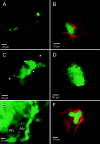
Figure 1.
Intravascular location of spontaneous lung metastases. 4T1 tumor cells that spontaneously metastasized into the lung were visualized by high-resolution digital video microscopy as described in Materials and Methods. In A, B, and F, the lung endothelium has been labeled with a red fluorescent dye, DiI-acetylated LDL. A: Solitary tumor cells (green) attached within the precapillary arterioles. B: Solitary tumor cell attached in a capillary. C: Colony of ∼10 cells within a blood vessel. The endothelial margins, indicated by arrows, are visible because of reflection and scattering of green fluorescent protein fluorescence from tumor cells. D: Large colony of tumor cells within the lung vasculature. E: Another large colony that probably has outgrown the vessel of initial attachment continues to exhibit intravascular growth along capillaries around alveoli (Alv). Note how the outline of the alveolar wall is formed by strings of green fluorescent protein-expressing tumor cells. F: Front projection of a three dimensional-reconstructed image of a small colony shows intravascular location. The endothelium was labeled with DiI-acetylated LDL (red). Fifty planes of a stack of images taken separately in the green and red fluorescence channels taken along the z axis spanning a depth of 50 μm were overlaid.
Similar articles
- Intravascular origin of metastasis from the proliferation of endothelium-attached tumor cells: a new model for metastasis.
Al-Mehdi AB, Tozawa K, Fisher AB, Shientag L, Lee A, Muschel RJ. Al-Mehdi AB, et al. Nat Med. 2000 Jan;6(1):100-2. doi: 10.1038/71429. Nat Med. 2000. PMID: 10613833 - The role of the intravascular microenvironment in spontaneous metastasis development.
Zhang Q, Yang M, Shen J, Gerhold LM, Hoffman RM, Xing HR. Zhang Q, et al. Int J Cancer. 2010 Jun 1;126(11):2534-41. doi: 10.1002/ijc.24979. Int J Cancer. 2010. PMID: 19847811 - Determination of clonality of metastasis by cell-specific color-coded fluorescent-protein imaging.
Yamamoto N, Yang M, Jiang P, Xu M, Tsuchiya H, Tomita K, Moossa AR, Hoffman RM. Yamamoto N, et al. Cancer Res. 2003 Nov 15;63(22):7785-90. Cancer Res. 2003. PMID: 14633704 - Cell detachment and metastasis.
Weiss L, Ward PM. Weiss L, et al. Cancer Metastasis Rev. 1983;2(2):111-27. doi: 10.1007/BF00048965. Cancer Metastasis Rev. 1983. PMID: 6352010 Review. - Tail vein assay of cancer metastasis.
Elkin M, Vlodavsky I. Elkin M, et al. Curr Protoc Cell Biol. 2001 Nov;Chapter 19:19.2.1-19.2.7. doi: 10.1002/0471143030.cb1902s12. Curr Protoc Cell Biol. 2001. PMID: 18228345 Review.
Cited by
- The role of immunoglobulin superfamily cell adhesion molecules in cancer metastasis.
Wai Wong C, Dye DE, Coombe DR. Wai Wong C, et al. Int J Cell Biol. 2012;2012:340296. doi: 10.1155/2012/340296. Epub 2012 Jan 9. Int J Cell Biol. 2012. PMID: 22272201 Free PMC article. - Dormant cancer cells retrieved from metastasis-free organs regain tumorigenic and metastatic potency.
Suzuki M, Mose ES, Montel V, Tarin D. Suzuki M, et al. Am J Pathol. 2006 Aug;169(2):673-81. doi: 10.2353/ajpath.2006.060053. Am J Pathol. 2006. PMID: 16877365 Free PMC article. - Inhibition of metastatic tumor formation in vivo by a bacteriophage display-derived galectin-3 targeting peptide.
Newton-Northup JR, Dickerson MT, Ma L, Besch-Williford CL, Deutscher SL. Newton-Northup JR, et al. Clin Exp Metastasis. 2013 Feb;30(2):119-32. doi: 10.1007/s10585-012-9516-y. Epub 2012 Aug 1. Clin Exp Metastasis. 2013. PMID: 22851004 - Lung endothelium exploits susceptible tumor cell states to instruct metastatic latency.
Jakab M, Lee KH, Uvarovskii A, Ovchinnikova S, Kulkarni SR, Jakab S, Rostalski T, Spegg C, Anders S, Augustin HG. Jakab M, et al. Nat Cancer. 2024 May;5(5):716-730. doi: 10.1038/s43018-023-00716-7. Epub 2024 Feb 2. Nat Cancer. 2024. PMID: 38308117 Free PMC article. - The two faces of transforming growth factor beta in carcinogenesis.
Roberts AB, Wakefield LM. Roberts AB, et al. Proc Natl Acad Sci U S A. 2003 Jul 22;100(15):8621-3. doi: 10.1073/pnas.1633291100. Epub 2003 Jul 14. Proc Natl Acad Sci U S A. 2003. PMID: 12861075 Free PMC article. Review. No abstract available.
References
- Al-Mehdi AB, Tozawa K, Fisher AB, Shientag L, Lee A, Muschel RJ: Intravascular origin of metastasis from proliferation of endothelium-attached tumor cells: a new model for metastasis. Nat Med 2000, 6:100-102 - PubMed
- Wong CW, Lee A, Shientag L, Yu J, Dong Y, Kao G, Al-Mehdi AB, Bernhard EJ, Muschel RJ: Apoptosis: an early event in metastatic inefficiency. Cancer Res 2001, 61:333-338 - PubMed
- Ito S, Nakanishi H, Ikehara Y, Kato T, Kasai Y, Ito K, Akiyama S, Nakao A, Tatematsu M: Real time observation of micrometastasis formation in the living mouse liver using a green fluorescent protein gene-tagged rat tongue carcinoma cell line. Int J Cancer , 93:212-217 - PubMed
- Aslakson CJ, Miller FR: Selective events in the metastatic process defined by analysis of the sequential dissemination of subpopulations of a mouse mammary tumor. Cancer Res 1992, 52:1399-1405 - PubMed
- Iwasaki T: Histological and experimental observations on the destruction of tumour cells in the blood vessels. J Pathol Bacteriol 1915, 20:85-105
Publication types
MeSH terms
Substances
LinkOut - more resources
Full Text Sources
Other Literature Sources
Medical
