Smaller hippocampal volume predicts pathologic vulnerability to psychological trauma - PubMed (original) (raw)
Smaller hippocampal volume predicts pathologic vulnerability to psychological trauma
Mark W Gilbertson et al. Nat Neurosci. 2002 Nov.
Abstract
In animals, exposure to severe stress can damage the hippocampus. Recent human studies show smaller hippocampal volume in individuals with the stress-related psychiatric condition posttraumatic stress disorder (PTSD). Does this represent the neurotoxic effect of trauma, or is smaller hippocampal volume a pre-existing condition that renders the brain more vulnerable to the development of pathological stress responses? In monozygotic twins discordant for trauma exposure, we found evidence that smaller hippocampi indeed constitute a risk factor for the development of stress-related psychopathology. Disorder severity in PTSD patients who were exposed to trauma was negatively correlated with the hippocampal volume of both the patients and the patients' trauma-unexposed identical co-twin. Furthermore, severe PTSD twin pairs-both the trauma-exposed and unexposed members-had significantly smaller hippocampi than non-PTSD pairs.
Conflict of interest statement
Competing interests statement
The authors declare that they have no competing financial interests.
Figures
Fig. 1
Discordant monozygotic twin paradigm for assessing MRI differences in PTSD. Sample coronal MRI images of right (red) and left (blue) hippocampi in a PTSD and a non-PTSD twin pair. Images represent four subject groups: (1) combat-exposed (Ex) subjects who developed chronic PTSD (ExP+); (2) their combat-unexposed (Ux) co-twins with no PTSD themselves (UxP+); (3) Ex subjects who never developed PTSD (ExP−) and (4) Ux co-twins also with no PTSD (UxP−). Contrast (a) provides a replication of previous work demonstrating smaller hippocampal volumes in combat veterans with versus without PTSD. Contrast (b) identifies the neurotoxicity effect—hippocampal reduction—as environmentally acquired, by contrasting hippocampal volumes in combat-exposed PTSD veterans with their unexposed co-twins. Contrast (c) examines pre-existing vulnerability by contrasting hippocampal volumes in the two groups of combat-unexposed co-twins whose combat-exposed brothers did versus did not develop PTSD. Model is tested by a diagnosis (P+ versus P−) × exposure (Ex versus Ux) ANOVA. Diagnosis refers to combat-exposed twin only. If hippocampal volume represents a vulnerability factor, the model predicts a significant main effect of diagnosis in the absence of a diagnosis × exposure interaction (that is, PTSD combat-exposed veterans and their unexposed co-twins show the same pattern). If hippocampal reduction results from neurotoxicity, the model predicts a significant main effect of exposure and/or a significant diagnosis × exposure interaction.
Fig. 2
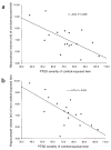
Hippocampal volume correlations with post-trauma symptoms. Scatter plots illustrate relationship of symptom severity in combat veterans with PTSD to: (a) their own hippocampal volumes and (b) the hippocampal volumes of their identical twin brothers who were not exposed to combat. Symptom severity represents the total score received on the Clinician-Administered PTSD Scale (CAPS).
Fig. 3
Total hippocampal volumes for four subject groups. Scatter plot illustrates absolute hippocampal volumes (ml) for combat-exposed individuals with and without PTSD, as well as for their respective unexposed co-twins. Data are only presented for PTSD twin pairs in which the combat-exposed twin had a CAPS score >65.
Comment in
- Chickens, eggs and hippocampal atrophy.
Sapolsky RM. Sapolsky RM. Nat Neurosci. 2002 Nov;5(11):1111-3. doi: 10.1038/nn1102-1111. Nat Neurosci. 2002. PMID: 12404003 Review. No abstract available.
Similar articles
- Configural cue performance in identical twins discordant for posttraumatic stress disorder: theoretical implications for the role of hippocampal function.
Gilbertson MW, Williston SK, Paulus LA, Lasko NB, Gurvits TV, Shenton ME, Pitman RK, Orr SP. Gilbertson MW, et al. Biol Psychiatry. 2007 Sep 1;62(5):513-20. doi: 10.1016/j.biopsych.2006.12.023. Epub 2007 May 23. Biol Psychiatry. 2007. PMID: 17509537 Free PMC article. - Subtle neurologic compromise as a vulnerability factor for combat-related posttraumatic stress disorder: results of a twin study.
Gurvits TV, Metzger LJ, Lasko NB, Cannistraro PA, Tarhan AS, Gilbertson MW, Orr SP, Charbonneau AM, Wedig MM, Pitman RK. Gurvits TV, et al. Arch Gen Psychiatry. 2006 May;63(5):571-6. doi: 10.1001/archpsyc.63.5.571. Arch Gen Psychiatry. 2006. PMID: 16651514 - Hippocampal volume, PTSD, and alcoholism in combat veterans.
Woodward SH, Kaloupek DG, Streeter CC, Kimble MO, Reiss AL, Eliez S, Wald LL, Renshaw PF, Frederick BB, Lane B, Sheikh JI, Stegman WK, Kutter CJ, Stewart LP, Prestel RS, Arsenault NJ. Woodward SH, et al. Am J Psychiatry. 2006 Apr;163(4):674-81. doi: 10.1176/ajp.2006.163.4.674. Am J Psychiatry. 2006. PMID: 16585443 - Hippocampal volume deficits associated with exposure to psychological trauma and posttraumatic stress disorder in adults: a meta-analysis.
Woon FL, Sood S, Hedges DW. Woon FL, et al. Prog Neuropsychopharmacol Biol Psychiatry. 2010 Oct 1;34(7):1181-8. doi: 10.1016/j.pnpbp.2010.06.016. Epub 2010 Jun 21. Prog Neuropsychopharmacol Biol Psychiatry. 2010. PMID: 20600466 Review. - Searching for non-genetic molecular and imaging PTSD risk and resilience markers: Systematic review of literature and design of the German Armed Forces PTSD biomarker study.
Schmidt U, Willmund GD, Holsboer F, Wotjak CT, Gallinat J, Kowalski JT, Zimmermann P. Schmidt U, et al. Psychoneuroendocrinology. 2015 Jan;51:444-58. doi: 10.1016/j.psyneuen.2014.08.020. Epub 2014 Sep 4. Psychoneuroendocrinology. 2015. PMID: 25236294 Review.
Cited by
- How Psychedelics Modulate Multiple Memory Mechanisms in Posttraumatic Stress Disorder.
Doss MK, DeMarco A, Dunsmoor JE, Cisler JM, Fonzo GA, Nemeroff CB. Doss MK, et al. Drugs. 2024 Oct 26. doi: 10.1007/s40265-024-02106-4. Online ahead of print. Drugs. 2024. PMID: 39455547 Review. - Borderline personality disorder and post-traumatic stress disorder in adolescents: protocol for a comparative study of borderline personality disorder with and without comorbid post-traumatic stress disorder (BORDERSTRESS-ADO).
Riou M, Duclos H, Leribillard M, Parienti JJ, Segobin S, Viard A, Apter G, Gerardin P, Guillery B, Guénolé F. Riou M, et al. BMC Psychiatry. 2024 Oct 23;24(1):724. doi: 10.1186/s12888-024-06093-4. BMC Psychiatry. 2024. PMID: 39443885 Free PMC article. - BMP Antagonist Gremlin 2 Regulates Hippocampal Neurogenesis and Is Associated with Seizure Susceptibility and Anxiety.
Frazer NB, Kaas GA, Firmin CG, Gamazon ER, Hatzopoulos AK. Frazer NB, et al. eNeuro. 2024 Oct 17;11(10):ENEURO.0213-23.2024. doi: 10.1523/ENEURO.0213-23.2024. Print 2024 Oct. eNeuro. 2024. PMID: 39349059 Free PMC article. - Impact of Childhood Maltreatment on Cognitive Function and Its Relationship With Emotion Regulation in Young Adults.
Kim MS, Kim K, Nam J, Lee SJ, Lee SW. Kim MS, et al. Soa Chongsonyon Chongsin Uihak. 2024 Jul 1;35(3):155-162. doi: 10.5765/jkacap.240001. Soa Chongsonyon Chongsin Uihak. 2024. PMID: 38966202 Free PMC article. - The Neglected Sibling: NLRP2 Inflammasome in the Nervous System.
Ducza L, Gaál B. Ducza L, et al. Aging Dis. 2024 May 7;15(3):1006-1028. doi: 10.14336/AD.2023.0926-1. Aging Dis. 2024. PMID: 38722788 Free PMC article. Review.
References
- McEwen BS. In: The Cognitive Neurosciences. Gazzaniga MS, editor. MIT Press; Cambridge, Massachusetts: 1995. pp. 1117–1135.
- Squire LR. Memory and the hippocampus: a synthesis from findings with rats, monkeys, and humans. Psychol Rev. 1992;99:195–231. - PubMed
- Zola-Morgan S, Squire LR. Neuroanatomy of memory. Annu Rev Neurosci. 1993;16:547–563. - PubMed
Publication types
MeSH terms
Grants and funding
- K02 MH001110-10/MH/NIMH NIH HHS/United States
- R01 MH054636/MH/NIMH NIH HHS/United States
- K02-MH01110/MH/NIMH NIH HHS/United States
- R01-MH54636/MH/NIMH NIH HHS/United States
LinkOut - more resources
Full Text Sources
Other Literature Sources
Medical


