Functional replacement of the carboxy-terminal two-thirds of the influenza A virus NS1 protein with short heterologous dimerization domains - PubMed (original) (raw)
Functional replacement of the carboxy-terminal two-thirds of the influenza A virus NS1 protein with short heterologous dimerization domains
Xiuyan Wang et al. J Virol. 2002 Dec.
Abstract
The NS1 protein of influenza A/WSN/33 virus is a 230-amino-acid-long protein which functions as an interferon alpha/beta (IFN-alpha/beta) antagonist by preventing the synthesis of IFN during viral infection. In tissue culture, the IFN inhibitory function of the NS1 protein has been mapped to the RNA binding domain, the first 73 amino acids. Nevertheless, influenza viruses expressing carboxy-terminally truncated NS1 proteins are attenuated in mice. Dimerization of the NS1 protein has previously been shown to be essential for its RNA binding activity. We have explored the ability of heterologous dimerization domains to functionally substitute in vivo for the carboxy-terminal domains of the NS1 protein. Recombinant influenza viruses were generated that expressed truncated NS1 proteins of 126 amino acids, fused to 28 or 24 amino acids derived from the dimerization domains of either the Saccharomyces cerevisiae PUT3 or the Drosophila melanogaster Ncd (DmNcd) proteins. These viruses regained virulence and lethality in mice. Moreover, a recombinant influenza virus expressing only the first 73 amino acids of the NS1 protein was able to replicate in mice lacking three IFN-regulated antiviral enzymes, PKR, RNaseL, and Mx, but not in wild-type (Mx-deficient) mice, suggesting that the attenuation was mainly due to an inability to inhibit the IFN system. Remarkably, a virus with an NS1 truncated at amino acid 73 but fused to the dimerization domain of DmNcd replicated and was also highly pathogenic in wild-type mice. These results suggest that the main biological function of the carboxy-terminal region of the NS1 protein of influenza A virus is the enhancement of its IFN antagonist properties by stabilizing the NS1 dimeric structure.
Figures
FIG. 1.
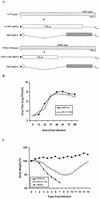
Wild-type influenza A/WSN/33 virus and recombinant WSN-NS1(1-126) virus. (A) Schematic representation of the NS genes and gene transcripts for wild-type (wt) influenza WSN virus and recombinant WSN-NS1(1-126) virus. Viral NS genes are indicated by light gray boxes, with nucleotide (nt) length indicated in numbers below the gene segments. Stop codons for wild-type NS1 and NS1(1-126) open reading frames are indicated by an asterisk above the viral genes. Viral nuclear export protein (NEP) mRNAs are also shown, with spotted boxes representing the specific open reading frames of the viral NEP mRNA transcripts. (B) Growth curves of WSN-NS1(1-126) and WSN viruses on MDBK cells. Confluent cell monolayers in 35-mm-diameter dishes were infected with wild-type influenza A/WSN/33 virus and with the mutant WSN-NS1(1-126) virus at an MOI of 0.001. At the indicated time points, infectious particles present in the media were titrated by plaque assay in MDBK cells. (C) Pathogenicity of WSN-NS1(1-126) virus in mice. Groups of five mice were infected intranasally with either wild-type WSN virus or WSN-NS1(1-126) virus at a dose of 2 × 10⁴ PFU/mouse. In addition, four mice were infected with 2 × 10⁴ PFU of delNS1 virus/mouse. Animals were weighed dailyfollowing infection. The body weights on each day postinfection are given as the percentage of the original body weight (on day 0). The average body weight percentages of animals per group are represented. aa, amino acid.
FIG. 2.
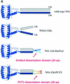
WSN-NS1(1-126)PUT3 and WSN-NS1(1-126)DmNcd viruses. (A) Schematic representation of the dimeric structures of the wild-type and mutant NS1(1-126), NS1(1-126)DmNcd, and NS1(1-126)PUT3 proteins. The crystal structure of the first 73 amino acids (aa) of the NS1 protein is adapted from reference . The crystal structure of the dimerization domain of DmNcd is adapted from reference . The structure of the dimerization domain of PUT3 is adapted from reference . The unknown structures are indicated by boxes. (B) Schematic representation of the NS genes and gene transcripts for influenza A WSN-NS1(1-126) dimerization domain-containing viruses, WSN-NS1(1-126)DmNcd and WSN-NS1(1-126)PUT3. NS genes and mRNA transcripts of the top two viruses are schematically represented as in Fig. 1. The 6-glycine linker region in WSN-NS1(1-126)DmNcd and WSN-NS1(1-126)PUT3 viruses is indicated by the striped box. The 24-amino-acid-long and 28-amino-acid-long dimerization domains from the D. melanogaster DmNcd protein or the S. cerevisiae PUT3 protein are represented by the checkered boxes. NEP, nuclear export protein; aa, amino acid; nt, nucleotide. (C) Western blot analysis of wild-type and mutant NS1 proteins in infected cells. MDBK cells were mock infected (mock) or were infected with wild-type (wt) WSN virus, recombinant WSN-NS1(1-126), WSN-NS1(1-126)DmNcd, or WSN-NS1(1-126)PUT3 virus at an MOI of 1. Sixteen hours postinfection, cell extracts were made and subjected to Western blot analysis with antibodies against the viral NP and NS1 proteins. (D) Pulse-chase analysis of wild-type NS1 and mutant NS1 proteins. MDBK cells were either mock infected or infected with wild-type WSN virus, WSN-NS1(1-126), WSN-NS1(1-126)DmNcd, or WSN-NS1(1-126)PUT3 virus at an MOI of 2. Four hours postinfection, cells were starved for methionine and cysteine and then pulse-labeled with 35S-Met and 35S-Cys for 30 min. At 1, 2, 4, and 20 h postchase total cell extracts were immunoprecipitated with antibodies against influenza NP and NS1 proteins. Samples were then boiled in SDS loading buffer and were separated by SDS-12% PAGE. The gel was then dried and subjected to autoradiography. h.p.c., hours postchase. (E) Pathogenicity of WSN-NS1(1-126)DmNcd and WSN-NS1(1-126)PUT3 viruses in mice. Groups of five mice were intranasally infected with 2 × 10⁴ PFU of either wild-type WSN, WSN-NS1(1-126), WSN-NS1(1-126)DmNcd, or WSN-NS1(1-126)PUT3 virus/mouse. Animals were inspected daily following infection. The percentage of surviving animals as a function of time is represented.
FIG. 3.
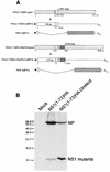
WSN-NS1(1-73)HA and WSN-NS1(1-73)HA-DmNcd viruses. (A) Schematic representation of the NS genes and gene transcripts for influenza A WSN-NS1(1-73)HA and WSN-NS1(1-73)HA-DmNcd viruses. Viral NS genes and mRNA transcripts are schematically represented as in Fig. 1. A deletion of 159 nucleotides (nt) in the NS1(1-73)HA gene is indicated by a vertical bar. A 9-amino-acid (aa) HA tag in WSN-NS1(1-73)HA and WSN-NS1(1-73)HA-DmNcd viral NS genes is presented by the dotted bar. The 6-glycine linker region in WSN-NS1(1-73)DmNcd virus is indicated by the striped box. The dimerization domain is represented by the checkered boxes as in Fig. 2. NEP, nuclear export protein. (B) Western blot analysis of mutant NS1 proteins in infected cells. MDBK cells were either mock infected (mock) or were infected with WSN-NS1(1-73)HA or WSN-NS1(1-73)HA-DmNcd virus at an MOI of 1. Sixteen hours postinfection, cell extracts were made and were subjected to Western blot analysis with antibodies against the viral NP protein and the HA tag. It should be noted that at the electrophoretic conditions used (SDS-12% PAGE), both NS1(1-73)HA and NS1(1-73)HA-DmNcd run with the front and do not show significant differences in electrophoretic mobility. Numbers at the left are molecular sizes.
FIG. 4.
Pathogenicity of WSN-NS1(1-73)HA and WSN-NS1(1-73)HA-DmNcd viruses in mice. Groups of five C57BL/6 mice were intranasally infected with 5 × 10⁵, 3.3 × 10⁴, or 1 × 10³ PFU of either WSN-NS1(1-73)HA or WSN-NS1(1-73)HA-DmNcd virus. Animals were weighed daily following infection, and body weights were compared with those from the day of infection. The average body weight percentages of animals per group are represented.
FIG. 5.
Dimerization and/or multimerization of wild-type NS1 and mutant NS1(1-73)HA and NS1(1-73)HA-DmNcd proteins, as measured by using a mammalian two-hybrid assay. 293T cells were cotransfected with pG5-Luc, pRL-TK, and Gal4-WSN-NS1 and VP16-WSN-NS1 (NS1, first column), Gal4-NS1(1-73)HA and VP16-NS1(1-73)HA [NS1(1-73)HA, second column], or Gal4-NS1(1-73)HA-DmNcd and VP16-NS1(1-73)HA-DmNcd plasmids [NS1(1-73)HA-DmNcd, third column]. Forty-eight hours posttransfection, cell extracts were made and luciferase activity was determined.
FIG. 6.
IFN-β gene induction by viruses expressing wild-type NS1 protein and mutant NS1 proteins. A549 cells were either mock infected (mock) or were infected with wild-type PR8, delNS1, wild-type WSN, WSN-NS1(1-126), WSN-NS1(1-126)DmNcd, WSN-NS1(1-126)PUT3, WSN-NS1(1-73)HA, or WSN-NS1(1-73)HA-DmNcd virus at an MOI of 1. At 10 and 20 h postinfection RNAs were extracted, and 10 μg of total RNAs from each sample was used for Northern blot analysis by using probes specific for the human IFN-β gene. As a control, 28S and 18S rRNAs were visualized by ethidium bromide staining.
Similar articles
- A recombinant influenza A virus expressing an RNA-binding-defective NS1 protein induces high levels of beta interferon and is attenuated in mice.
Donelan NR, Basler CF, García-Sastre A. Donelan NR, et al. J Virol. 2003 Dec;77(24):13257-66. doi: 10.1128/jvi.77.24.13257-13266.2003. J Virol. 2003. PMID: 14645582 Free PMC article. - The N- and C-terminal domains of the NS1 protein of influenza B virus can independently inhibit IRF-3 and beta interferon promoter activation.
Donelan NR, Dauber B, Wang X, Basler CF, Wolff T, García-Sastre A. Donelan NR, et al. J Virol. 2004 Nov;78(21):11574-82. doi: 10.1128/JVI.78.21.11574-11582.2004. J Virol. 2004. PMID: 15479798 Free PMC article. - Mutations in the NS1 protein of swine influenza virus impair anti-interferon activity and confer attenuation in pigs.
Solórzano A, Webby RJ, Lager KM, Janke BH, García-Sastre A, Richt JA. Solórzano A, et al. J Virol. 2005 Jun;79(12):7535-43. doi: 10.1128/JVI.79.12.7535-7543.2005. J Virol. 2005. PMID: 15919908 Free PMC article. - Attenuated influenza virus vaccines with modified NS1 proteins.
Richt JA, García-Sastre A. Richt JA, et al. Curr Top Microbiol Immunol. 2009;333:177-95. doi: 10.1007/978-3-540-92165-3_9. Curr Top Microbiol Immunol. 2009. PMID: 19768406 Review. - The influenza virus NS1 protein: inhibitor of innate and adaptive immunity.
Fernandez-Sesma A. Fernandez-Sesma A. Infect Disord Drug Targets. 2007 Dec;7(4):336-43. doi: 10.2174/187152607783018754. Infect Disord Drug Targets. 2007. PMID: 18220965 Review.
Cited by
- Antiviral responses versus virus-induced cellular shutoff: a game of thrones between influenza A virus NS1 and SARS-CoV-2 Nsp1.
Khalil AM, Nogales A, Martínez-Sobrido L, Mostafa A. Khalil AM, et al. Front Cell Infect Microbiol. 2024 Feb 5;14:1357866. doi: 10.3389/fcimb.2024.1357866. eCollection 2024. Front Cell Infect Microbiol. 2024. PMID: 38375361 Free PMC article. Review. - Interaction between NS1 and Cellular MAVS Contributes to NS1 Mitochondria Targeting.
Tseng YY, Kuan CY, Mibayashi M, Chen CJ, Palese P, Albrecht RA, Hsu WL. Tseng YY, et al. Viruses. 2021 Sep 23;13(10):1909. doi: 10.3390/v13101909. Viruses. 2021. PMID: 34696339 Free PMC article. - Non-structural protein 1 of avian influenza A viruses differentially inhibit NF-κB promoter activation.
Munir M, Zohari S, Berg M. Munir M, et al. Virol J. 2011 Aug 2;8:383. doi: 10.1186/1743-422X-8-383. Virol J. 2011. PMID: 21810221 Free PMC article. - A transient homotypic interaction model for the influenza A virus NS1 protein effector domain.
Kerry PS, Ayllon J, Taylor MA, Hass C, Lewis A, García-Sastre A, Randall RE, Hale BG, Russell RJ. Kerry PS, et al. PLoS One. 2011 Mar 28;6(3):e17946. doi: 10.1371/journal.pone.0017946. PLoS One. 2011. PMID: 21464929 Free PMC article. - The R35 residue of the influenza A virus NS1 protein has minimal effects on nuclear localization but alters virus replication through disrupting protein dimerization.
Lalime EN, Pekosz A. Lalime EN, et al. Virology. 2014 Jun;458-459:33-42. doi: 10.1016/j.virol.2014.04.012. Epub 2014 May 5. Virology. 2014. PMID: 24928037 Free PMC article.
References
Publication types
MeSH terms
Substances
LinkOut - more resources
Full Text Sources
Other Literature Sources
Molecular Biology Databases


