CD4+CD25+ T(R) cells suppress innate immune pathology through cytokine-dependent mechanisms - PubMed (original) (raw)
CD4+CD25+ T(R) cells suppress innate immune pathology through cytokine-dependent mechanisms
Kevin J Maloy et al. J Exp Med. 2003.
Abstract
CD4(+)CD25(+) regulatory T (T(R)) cells can inhibit a variety of autoimmune and inflammatory diseases, but the precise mechanisms by which they suppress immune responses in vivo remain unresolved. Here, we have used Helicobacter hepaticus infection of T cell-reconstituted recombination-activating gene (RAG)(-/-) mice as a model to study the ability of CD4(+)CD25(+) T(R) cells to inhibit bacterially triggered intestinal inflammation. H. hepaticus infection elicited both T cell-mediated and T cell-independent intestinal inflammation, both of which were inhibited by adoptively transferred CD4(+)CD25(+) T(R) cells. T cell-independent pathology was accompanied by activation of the innate immune system that was also inhibited by CD4(+)CD25(+) T(R) cells. Suppression of innate immune pathology was dependent on T cell-derived interleukin 10 and also on the production of transforming growth factor beta. Thus, CD4(+)CD25(+) T(R) cells do not only suppress adaptive T cell responses, but are also able to control pathology mediated by innate immune mechanisms.
Figures
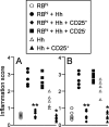
Figure 1.
CD4+CD25+ T cells prevent both T cell–dependent and T cell–independent intestinal inflammation. 129SvEvRAG2−/− mice were reconstituted with purified CD4+ T cell subsets as indicated (4 × 10⁵ CD45RBhigh T cells and/or 105 CD4+CD45RBlowCD25+ T cells or CD4+CD45RBlowCD25− T cells) and infected 24 h later by oral gavage with 2 × 10⁸ cfu H. hepaticus. Mice were killed 8–12 wk later and pathology in the colon (A) and cecum (B) was assessed histologically. Each symbol represents a single animal and results are representative of two similar experiments. **P < 0.01 vs. RBhi + Hh; *P < 0.05 vs. Hh.
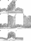
Figure 2.
Representative photomicrographs of hematoxylin and eosin stained intestinal tissues from 129SvEvRAG2−/− mice that were reconstituted with purified CD4+ T cell subsets and/or infected with H. hepaticus as outlined in Fig. 1. (A–G) Colon sections from a control RAG2−/− mouse (A) and from mice that received; H. hepaticus only (B), H. hepaticus and CD4+CD45RBlowCD25+ T cells (C), CD4+CD45RBhigh T cells only (D), CD4+CD45RBhigh T cells and H. hepaticus (E), CD45RBhighCD4+ T cells plus CD4+CD45RBlowCD25+ T cells and H. hepaticus (F), or CD45RBhighCD4+ T cells plus CD4+CD45RBlowCD25− T cells and H. hepaticus (G). (H–J) Cecum sections from a control RAG2−/− mouse (H) and from mice that received; H. hepaticus only (I), or H. hepaticus and CD4+CD45RBlowCD25+ T cells (J). Original magnification: ×50.
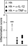
Figure 3.
_H. hepaticus_–triggered T cell–independent intestinal inflammation is driven by proinflammatory cytokines. 129SvEvRAG2−/− mice were infected by oral gavage with 2 × 10⁸ cfu H. hepaticus. Where indicated, mice received 1 mg/wk anti–IL-12p40 for the first 2 wk after infection or 2 mg/wk anti-TNF-α throughout the course of the infection. Mice were killed 8–12 wk later and pathology in the cecum was assessed histologically. Each symbol represents a single animal and results are representative of two similar experiments. **P < 0.01 vs. anti–IL-12p40 or anti-TNF-α.
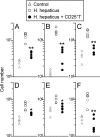
Figure 4.
H. hepaticus infection of RAG2−/− mice induces systemic activation of innate immunity that is inhibited by CD4+CD25+ T cells. 129SvEvRAG2−/− mice were reconstituted with 105 CD4+ CD45RBlowCD25+ T cells and/or infected by oral gavage with 2 × 10⁸ cfu H. hepaticus. Mice were killed 8–12 wk later and spleen cell populations were enumerated using FACS® analyses. Graphs shown represent numbers of total spleen cells (A), neutrophils (B), monocytes/macrophages (C), dendritic cells (D), NK cells (E), and IFN-γ–producing NK cells (F). Each symbol represents a single animal and results are representative of two similar experiments. **P < 0.01 vs. Hh; *P < 0.05 vs. Hh.
Figure 5.
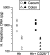
CD4+CD25+ T cell reconstitution does not affect H. hepaticus colonization levels in RAG2−/− mice. 129SvEvRAG2−/− mice were reconstituted with 105 CD4+ CD45RBlowCD25+ T cells and/or infected by oral gavage with 2 × 10⁸ cfu H. hepaticus. Mice were killed 10 wk later, DNA was isolated from cecum and colon samples and H. hepaticus DNA was quantified using a real-time PCR assay. Each symbol represents a single animal and results are representative of two similar experiments.
Figure 6.
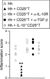
CD4+CD25+ T cell–mediated protection against T cell–independent intestinal inflammation is dependent on IL-10 and TGF-β. 129SvEvRAG2−/− mice were reconstituted with 105 CD4+CD45RBlowCD25+ T cells from wild-type or IL-10−/− mice and/or infected by oral gavage with 2 × 10⁸ cfu H. hepaticus. Where indicated mice received 0.5 mg/wk anti–IL-10R or 2 mg/wk anti-TGF-β. Mice were killed 8–12 wk later, ceca samples were taken for histological examination. Each symbol represents a single animal and results are representative of two similar experiments. **P < 0.01 vs. Hh.
Similar articles
- Cure of innate intestinal immune pathology by CD4+CD25+ regulatory T cells.
Maloy KJ, Antonelli LR, Lefevre M, Powrie F. Maloy KJ, et al. Immunol Lett. 2005 Mar 15;97(2):189-92. doi: 10.1016/j.imlet.2005.01.004. Immunol Lett. 2005. PMID: 15752557 Review. - Bacteria-triggered CD4(+) T regulatory cells suppress Helicobacter hepaticus-induced colitis.
Kullberg MC, Jankovic D, Gorelick PL, Caspar P, Letterio JJ, Cheever AW, Sher A. Kullberg MC, et al. J Exp Med. 2002 Aug 19;196(4):505-15. doi: 10.1084/jem.20020556. J Exp Med. 2002. PMID: 12186842 Free PMC article. - CD4+ CD25+ regulatory T lymphocytes inhibit microbially induced colon cancer in Rag2-deficient mice.
Erdman SE, Poutahidis T, Tomczak M, Rogers AB, Cormier K, Plank B, Horwitz BH, Fox JG. Erdman SE, et al. Am J Pathol. 2003 Feb;162(2):691-702. doi: 10.1016/S0002-9440(10)63863-1. Am J Pathol. 2003. PMID: 12547727 Free PMC article. - CD4(+)CD25(+) regulatory lymphocytes require interleukin 10 to interrupt colon carcinogenesis in mice.
Erdman SE, Rao VP, Poutahidis T, Ihrig MM, Ge Z, Feng Y, Tomczak M, Rogers AB, Horwitz BH, Fox JG. Erdman SE, et al. Cancer Res. 2003 Sep 15;63(18):6042-50. Cancer Res. 2003. PMID: 14522933 - Control of immune pathology by regulatory T cells.
Powrie F, Read S, Mottet C, Uhlig H, Maloy K. Powrie F, et al. Novartis Found Symp. 2003;252:92-8; discussion 98-105, 106-14. Novartis Found Symp. 2003. PMID: 14609214 Review.
Cited by
- Regulatory T cells: present facts and future hopes.
Becker C, Stoll S, Bopp T, Schmitt E, Jonuleit H. Becker C, et al. Med Microbiol Immunol. 2006 Sep;195(3):113-24. doi: 10.1007/s00430-006-0017-y. Epub 2006 May 20. Med Microbiol Immunol. 2006. PMID: 16715254 Review. - Interleukin-23 drives innate and T cell-mediated intestinal inflammation.
Hue S, Ahern P, Buonocore S, Kullberg MC, Cua DJ, McKenzie BS, Powrie F, Maloy KJ. Hue S, et al. J Exp Med. 2006 Oct 30;203(11):2473-83. doi: 10.1084/jem.20061099. Epub 2006 Oct 9. J Exp Med. 2006. PMID: 17030949 Free PMC article. - Innate lymphoid cells drive interleukin-23-dependent innate intestinal pathology.
Buonocore S, Ahern PP, Uhlig HH, Ivanov II, Littman DR, Maloy KJ, Powrie F. Buonocore S, et al. Nature. 2010 Apr 29;464(7293):1371-5. doi: 10.1038/nature08949. Nature. 2010. PMID: 20393462 Free PMC article. - Heme oxygenase-1 attenuates ovalbumin-induced airway inflammation by up-regulation of foxp3 T-regulatory cells, interleukin-10, and membrane-bound transforming growth factor- 1.
Xia ZW, Xu LQ, Zhong WW, Wei JJ, Li NL, Shao J, Li YZ, Yu SC, Zhang ZL. Xia ZW, et al. Am J Pathol. 2007 Dec;171(6):1904-14. doi: 10.2353/ajpath.2007.070096. Epub 2007 Nov 8. Am J Pathol. 2007. PMID: 17991714 Free PMC article. - Harnessing regulatory T cells to suppress asthma: from potential to therapy.
Thorburn AN, Hansbro PM. Thorburn AN, et al. Am J Respir Cell Mol Biol. 2010 Nov;43(5):511-9. doi: 10.1165/rcmb.2009-0342TR. Epub 2010 Jan 22. Am J Respir Cell Mol Biol. 2010. PMID: 20097830 Free PMC article. Review.
References
- Strober, W., I.J. Fuss, and R.S. Blumberg. 2002. The immunology of mucosal models of inflammation. Annu. Rev. Immunol. 20:495–549. - PubMed
- Khoo, U.Y., I.E. Proctor, and A.J. Macpherson. 1997. CD4+ T cell down-regulation in human intestinal mucosa: evidence for intestinal tolerance to luminal bacterial antigens. J. Immunol. 158:3626–3634. - PubMed
- Powrie, F., M.W. Leach, S. Mauze, L.B. Caddle, and R.L. Coffman. 1993. Phenotypically distinct subsets of CD4+ T cells induce or protect from chronic intestinal inflammation in C. B-17 scid mice. Int. Immunol. 5:1461–1471. - PubMed
- Morrissey, P.J., K. Charrier, S. Braddy, D. Liggitt, and J.D. Watson. 1993. CD4+ T cells that express high levels of CD45RB induce wasting disease when transferred into congenic severe combined immunodeficient mice. Disease development is prevented by cotransfer of purified CD4+ T cells. J. Exp. Med. 178:237–244. - PMC - PubMed
Publication types
MeSH terms
Substances
LinkOut - more resources
Full Text Sources
Other Literature Sources
Medical
Research Materials
