CEACAM1-4S, a cell-cell adhesion molecule, mediates apoptosis and reverts mammary carcinoma cells to a normal morphogenic phenotype in a 3D culture - PubMed (original) (raw)
CEACAM1-4S, a cell-cell adhesion molecule, mediates apoptosis and reverts mammary carcinoma cells to a normal morphogenic phenotype in a 3D culture
Julia Kirshner et al. Proc Natl Acad Sci U S A. 2003.
Abstract
In a 3D model of breast morphogenesis, CEACAM1 (carcinoembryonic antigen-related cell adhesion molecule 1) plays an essential role in lumen formation in a subline of the nonmalignant human breast cell line (MCF10A). We show that mammary carcinoma cells (MCF7), which do not express CEACAM1 or form lumena when grown in Matrigel, are restored to a normal morphogenic program when transfected with CEACAM1-4S, the short cytoplasmic isoform of CEACAM1 that predominates in breast epithelia. During the time course of lumen formation, CEACAM1-4S was found initially between the cells, and in mature acini, it was found exclusively in an apical location, identical to its expression pattern in normal breast. Lumena were formed by apoptosis as opposed to necrosis of the central cells within the alveolar structures, and apoptotic cells within the lumena expressed CEACAM1-4S. Dying cells exhibited classical hallmarks of apoptosis, including nuclear condensation, membrane blebbing, caspase activation, and DNA laddering. Apoptosis was mediated by Bax translocation to the mitochondria and release of cytochrome c into the cytoplasm, and was partially inhibited by culturing cells with caspase inhibitors. The dynamic changes in CEACAM1 expression during morphogenesis, together with studies implicating extracellular matrix and integrin signaling, suggest that a morphogenic program integrates cell-cell and cell-extracellular matrix signaling to produce the lumena in mammary glands. This report reveals a function of CEACAM1-4S relevant to cellular physiology that distinguishes it from its related long cytoplasmic domain isoform.
Figures
Figure 1
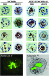
Time course of apoptosis and lumen formation in normal and carcinoma cells. MCF10F and MCF7/CEACAM1-4S cells (2.5 × 10⁵ per well) were grown in Matrigel for the indicated number of days, fixed, embedded in paraffin, and stained with caspase-cleaved cytokeratin 18 detecting mAb (M30) and anti-CEACAM1 mAb (4D1C2). For annexin V expression, cells were grown in Matrigel for 8 days, stained with annexin V-FITC, and visualized by confocal microscopy. For MCF7/CEACAM1-4S cells, the annexin V-FITC staining image was superimposed on the transmission image for easier visualization. (Magnification, ×600.)
Figure 2
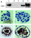
CEACAM1-4S mediated reversion of MCF7 cells to normal phenotype. (A) Cell lysate preparations (60 μg of total protein) from cells grown on plastic were separated by 8% reducing SDS/PAGE gel and immunoblotted with anti-CEACAM1 mAb (4D1C2). Lane 1, positive control HT29 colon carcinoma cells; lane 2, parental MCF7 cells; lane 3, MCF7/pHβ (vector); lane 4, MCF7/CEACAM1-4S; lane 5, MCF7/CEACAM1-4L. (B_–_E) Cells (2.5 × 10⁵ per well) were grown in Matrigel for 12 days, fixed, and embedded in paraffin, and sections were stained with anti-CEACAM1 mAb (4D1C2). (B) MCF7 cells exhibiting no lumen formation. (C) Vector-transfected MCF7 cells exhibiting no lumen formation. (D) Nonmalignant mammary epithelial cells MCF10F exhibiting lumen formation. (E) MCF7/CEACAM1-4S-transfected cells exhibiting lumen formation. (Magnification, ×600.)
Figure 3
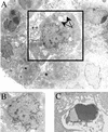
Morphologic analysis of apoptotic cells during lumen formation. MCF7/CEACAM1-4S cells (2.5 × 10⁵ per well) were grown in Matrigel for 5 (A and B) or 12 (C) days, and transmission electron microscopy was performed. (A) An acinus undergoing apoptosis of the central cell (box). (Magnification, ×5,500.) (B) Nuclear condensation occurring during apoptosis of the central cell seen in A. (Magnification, ×7,375.) (C) An apoptotic body in the lumen of the acinus. (Magnification, ×16,525.) Arrows point to the vacuoles seen in the cytoplasm of the apoptotic cell.
Figure 4
Mitochondrial potential and CEACAM1-4S expression in apoptotic cells. Cells (2.5 × 10⁵ per well) were grown in Matrigel for the indicated number of days, stained with JC-1 dye, and visualized by confocal microscopy. (A) MCF7/pHβ (vector) cells stained with JC-1 dye. (B) MCF7/CEACAM1-4S cells stained with JC-1 dye. (C) MCF7/CEACAM1-4S-ectoGFP cells. (D_–_F) MCF7/CEACAM1-4S-ectoGFP cells stained with JC-1 dye. (Magnification, ×600.)
Figure 5
Bax expression and cytochrome c release during lumen formation. (A) MCF7/pHβ (vector) or MCF7/CEACAM1-4S cells (2.5 × 10⁵ per well) were grown in Matrigel for 5 days, lysed, and fractionated into mitochondrial (M) and cytosolic (C) fractions. Total lysates (T) and the fractions of vector and CEACAM1-4S-transfected MCF7 cells were separated on an SDS/PAGE gel and immunoblotted with anti-Bax antibody. (B and C) Cells (2.5 × 10⁵ per well) were grown in Matrigel for 12 days, fixed, and embedded in paraffin, and sections were stained with anti-cytochrome c antibody. (B) MCF7/pHβ (vector). (C) MCF7/CEACAM1-4S. (Magnification, ×600.)
Similar articles
- Cell-cell adhesion molecule CEACAM1 is expressed in normal breast and milk and associates with beta1 integrin in a 3D model of morphogenesis.
Kirshner J, Hardy J, Wilczynski S, Shively JE. Kirshner J, et al. J Mol Histol. 2004 Mar;35(3):287-99. doi: 10.1023/b:hijo.0000032360.01976.81. J Mol Histol. 2004. PMID: 15339048 Free PMC article. Review. - Role of calpain-9 and PKC-delta in the apoptotic mechanism of lumen formation in CEACAM1 transfected breast epithelial cells.
Chen CJ, Nguyen T, Shively JE. Chen CJ, et al. Exp Cell Res. 2010 Feb 15;316(4):638-48. doi: 10.1016/j.yexcr.2009.11.001. Epub 2009 Nov 10. Exp Cell Res. 2010. PMID: 19909740 Free PMC article. - Mutational analysis of the cytoplasmic domain of CEACAM1-4L in humanized mammary glands reveals key residues involved in lumen formation: stimulation by Thr-457 and inhibition by Ser-461.
Li C, Chen CJ, Shively JE. Li C, et al. Exp Cell Res. 2009 Apr 15;315(7):1225-33. doi: 10.1016/j.yexcr.2008.12.015. Epub 2008 Dec 30. Exp Cell Res. 2009. PMID: 19146852 Free PMC article. - [CEACAM1--a less well-known member of the family of carcinoembryonic antigens].
Muchová L, Vítek L, Jirsa M. Muchová L, et al. Cas Lek Cesk. 2003;142(5):259-63. Cas Lek Cesk. 2003. PMID: 12920788 Review. Czech.
Cited by
- Calcium wave propagation in networks of endothelial cells: model-based theoretical and experimental study.
Long J, Junkin M, Wong PK, Hoying J, Deymier P. Long J, et al. PLoS Comput Biol. 2012;8(12):e1002847. doi: 10.1371/journal.pcbi.1002847. Epub 2012 Dec 27. PLoS Comput Biol. 2012. PMID: 23300426 Free PMC article. - Silica-based branched hollow microfibers as a biomimetic extracellular matrix for promoting tumor cell growth in vitro and in vivo.
Qiu P, Qu X, Brackett DJ, Lerner MR, Li D, Mao C. Qiu P, et al. Adv Mater. 2013 May 7;25(17):2492-6. doi: 10.1002/adma.201204472. Epub 2013 Mar 1. Adv Mater. 2013. PMID: 23450784 Free PMC article. - Rapid formation of size-controllable multicellular spheroids via 3D acoustic tweezers.
Chen K, Wu M, Guo F, Li P, Chan CY, Mao Z, Li S, Ren L, Zhang R, Huang TJ. Chen K, et al. Lab Chip. 2016 Jul 5;16(14):2636-43. doi: 10.1039/c6lc00444j. Lab Chip. 2016. PMID: 27327102 Free PMC article. - Prognostic Impact of CEACAM1 in Node-Negative Ovarian Cancer Patients.
Oliveira-Ferrer L, Goswami R, Galatenko V, Ding Y, Eylmann K, Legler K, Kürti S, Schmalfeldt B, Milde-Langosch K. Oliveira-Ferrer L, et al. Dis Markers. 2018 Jun 28;2018:6714287. doi: 10.1155/2018/6714287. eCollection 2018. Dis Markers. 2018. PMID: 30050594 Free PMC article. - Regulation of CEACAM1 transcription in human breast epithelial cells.
Gencheva M, Chen CJ, Nguyen T, Shively JE. Gencheva M, et al. BMC Mol Biol. 2010 Nov 4;11:79. doi: 10.1186/1471-2199-11-79. BMC Mol Biol. 2010. PMID: 21050451 Free PMC article.
References
- Svenberg T, Hammarstrom S, Hedin A. Mol Immunol. 1979;16:245–252. - PubMed
Publication types
MeSH terms
Substances
LinkOut - more resources
Full Text Sources
Medical
Molecular Biology Databases
Research Materials
Miscellaneous


