Contribution of transplanted bone marrow cells to Purkinje neurons in human adult brains - PubMed (original) (raw)
Contribution of transplanted bone marrow cells to Purkinje neurons in human adult brains
James M Weimann et al. Proc Natl Acad Sci U S A. 2003.
Abstract
We show here that cells within human adult bone marrow can contribute to cells in the adult human brain. Cerebellar tissues from female patients with hematologic malignancies, who had received chemotherapy, radiation, and a bone marrow transplant, were analyzed. Brain samples were obtained at autopsy from female patients who received male (sex-mismatched) or female (sex-matched, control) bone marrow transplants. Cerebella were evaluated in 10-microm-thick, formaldehyde-fixed, paraffin-embedded sections that encompassed up to approximately 50% of a human Purkinje nucleus. A total of 5,860 Purkinje cells from sex-mismatched females and 3,202 Purkinje cells from sex-matched females were screened for Y chromosomes by epifluorescence. Confocal laser scanning microscopy allowed definitive identification of the sex chromosomes within the morphologically distinct Purkinje cells. In the brains of females who received male bone marrow, four Purkinje neurons were found that contained an X and a Y chromosome and two other Purkinje neurons contained more than a diploid number of sex chromosomes. No Y chromosomes were detected in the brains of sex-matched controls. The total frequency of male bone marrow contribution to female Purkinje cells approximated 0.1%. This study demonstrates that although during human development Purkinje neurons are no longer generated after birth, cells within the bone marrow can contribute to these CNS neurons even in adulthood. The underlying mechanism may be caused either by generation de novo of Purkinje neurons from bone marrow-derived cells or by fusion of marrow-derived cells with existing recipient Purkinje neurons.
Figures
Figure 1
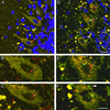
Controls. Identification of Purkinje neurons and specificity of the human X and Y chromosome DNA probes. Sections from control female and male cerebella were processed with a mixture of the X (red) and Y (green) probes. The nucleus was counterstained with To-Pro-3 (blue) and imaged by using a scanning confocal microscope at 1-μm optical sections. (A_–_C) Labeling in a female control cerebellum. (A) Two Purkinje neurons are clearly defined with a large nucleus surrounded by a large cytoplasmic region. Each cell has one X chromosome labeled (red arrows). (B and C) Enlargements of the Purkinje neurons in A without the blue nuclear label to facilitate visualization of the chromosome. Note that many of the granular neurons have two X chromosomes in contrast to the large Purkinje cells. (D_–_F) Male control cerebellum with three Purkinje neurons labeled. Note that one neuron has one X chromosome (red arrow), another has an X and a Y chromosome (red and green arrows), whereas the third has only one Y chromosome (green arrow). (E and F) Enlargements of D without the blue nuclear label. The white arrowhead in D and F shows a Y chromosome from a closely abutting cell. Note that many of the granular neurons contain one X (red) and one Y (green) chromosome. (Scale bars: 20 μm, A and D; 10 μm, B, C, E, and F.)
Figure 2
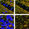
Male donor-derived cells in the circulation as well as the parenchyma of the cerebellum. (A) Donor-derived male blood cells are present in blood vessels in female recipients (field is representative of 20 examples imaged). (B) Same image as in A with the blue nuclear labeling removed. (C) Occasionally donor-derived cells can be found in the granular and molecular layers of the cerebellum possibly en route to the Purkinje layer (field is representative of >30 images captured). (D) Same image as in C with the blue nuclear labeling removed. (Scale bar: 10 μm.)
Figure 3
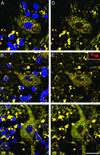
Evidence of male Y chromosome in Purkinje neurons. (A_–_C) Three examples of male bone marrow-derived nuclei in Purkinje cell. Each neuron has one X (red arrow) and one Y (green arrow) chromosome. These Purkinje neurons appear to be well integrated into the surrounding cerebellum with a mature morphology including dendrites. (D_–_F) Same images as A_–_C with the blue nuclear counterstain removed to highlight the red and green probes. The single X chromosome imaged in B and E has a dumbbell shape, a phenomenon observed infrequently. This chromosome is enlarged in the Inset (E) to demonstrate that it is a single chromosome. Note the wisp of red-labeled chromatin connecting the two lobes that are 1.18 μm apart, whereas the Y chromosome is 3.52 μm from the X. (Scale bar: 20 μm.)
Figure 4
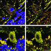
Evidence for fusion between donor-derived bone marrow cells and host Purkinje neurons. Two examples of triple sex chromosomes in Purkinje neurons. (A) Male to female transplant. There are two X (red) and one Y (green) chromosomes in this cell. (B) Same image as A without the nuclear counterstain. The two X chromosomes are 4.22 μm apart, whereas the Y chromosome is 4.44 μm and 6.08 μm separated from the two X chromosomes. The distance between each chromosome indicates that these are each unique chromosomes. (C) Male to female transplant. There are three distinct X chromosomes (red arrows) in this cell. These chromosomes are 4.16 μm, 6.12 μm, and 4.61 μm apart. (Scale bar: 10 μm.)
Similar articles
- Transplanted bone marrow generates new neurons in human brains.
Mezey E, Key S, Vogelsang G, Szalayova I, Lange GD, Crain B. Mezey E, et al. Proc Natl Acad Sci U S A. 2003 Feb 4;100(3):1364-9. doi: 10.1073/pnas.0336479100. Epub 2003 Jan 21. Proc Natl Acad Sci U S A. 2003. PMID: 12538864 Free PMC article. - Transplanted human bone marrow cells generate new brain cells.
Crain BJ, Tran SD, Mezey E. Crain BJ, et al. J Neurol Sci. 2005 Jun 15;233(1-2):121-3. doi: 10.1016/j.jns.2005.03.017. Epub 2005 Apr 21. J Neurol Sci. 2005. PMID: 15949500 Review. - Fusion of hematopoietic cells with Purkinje neurons does not lead to stable heterokaryon formation under noninvasive conditions.
Nern C, Wolff I, Macas J, von Randow J, Scharenberg C, Priller J, Momma S. Nern C, et al. J Neurosci. 2009 Mar 25;29(12):3799-807. doi: 10.1523/JNEUROSCI.5848-08.2009. J Neurosci. 2009. PMID: 19321776 Free PMC article. - Bone marrow transdifferentiation in brain after transplantation: a retrospective study.
Cogle CR, Yachnis AT, Laywell ED, Zander DS, Wingard JR, Steindler DA, Scott EW. Cogle CR, et al. Lancet. 2004 May 1;363(9419):1432-7. doi: 10.1016/S0140-6736(04)16102-3. Lancet. 2004. PMID: 15121406 - Binuclear Purkinje neurons.
Paltsyn AA, Komissarova SV. Paltsyn AA, et al. Patol Fiziol Eksp Ter. 2016 Oct-Dec;60(4):107-13. Patol Fiziol Eksp Ter. 2016. PMID: 29244931 Review.
Cited by
- Choice of Cell Source in Cell-Based Therapies for Retinal Damage due to Age-Related Macular Degeneration: A Review.
John S, Natarajan S, Parikumar P, Shanmugam P M, Senthilkumar R, Green DW, Abraham SJ. John S, et al. J Ophthalmol. 2013;2013:465169. doi: 10.1155/2013/465169. Epub 2013 Apr 22. J Ophthalmol. 2013. PMID: 23710332 Free PMC article. - Myelin repair: the role of stem and precursor cells in multiple sclerosis.
Chandran S, Hunt D, Joannides A, Zhao C, Compston A, Franklin RJ. Chandran S, et al. Philos Trans R Soc Lond B Biol Sci. 2008 Jan 12;363(1489):171-83. doi: 10.1098/rstb.2006.2019. Philos Trans R Soc Lond B Biol Sci. 2008. PMID: 17282989 Free PMC article. Review. - Membrane properties of neuron-like cells generated from adult human bone-marrow-derived mesenchymal stem cells.
Fox LE, Shen J, Ma K, Liu Q, Shi G, Pappas GD, Qu T, Cheng J. Fox LE, et al. Stem Cells Dev. 2010 Dec;19(12):1831-41. doi: 10.1089/scd.2010.0089. Epub 2010 Sep 13. Stem Cells Dev. 2010. PMID: 20394468 Free PMC article. - Fusion of proinsulin-producing bone marrow-derived cells with hepatocytes in diabetes.
Fujimiya M, Kojima H, Ichinose M, Arai R, Kimura H, Kashiwagi A, Chan L. Fujimiya M, et al. Proc Natl Acad Sci U S A. 2007 Mar 6;104(10):4030-5. doi: 10.1073/pnas.0700220104. Epub 2007 Feb 27. Proc Natl Acad Sci U S A. 2007. PMID: 17360472 Free PMC article. - Experimental neurotransplantation treatment for hereditary cerebellar ataxias.
Cendelin J. Cendelin J. Cerebellum Ataxias. 2016 Apr 4;3:7. doi: 10.1186/s40673-016-0045-3. eCollection 2016. Cerebellum Ataxias. 2016. PMID: 27047666 Free PMC article. Review.
References
- Korbling M, Katz R L, Khanna A, Ruifrok A C, Rondon G, Albitar M, Champlin R E, Estrov Z. N Engl J Med. 2002;346:738–746. - PubMed
- Okamoto R, Yajima T, Yamazaki M, Kanai T, Mukai M, Okamoto S, Ikeda Y, Hibi T, Inazawa J, Watanabe M. Nat Med. 2002;8:1011–1017. - PubMed
- Mezey E, Chandross K J, Harta G, Maki R A, McKercher S R. Science. 2000;290:1779–1782. - PubMed
- Brazelton T R, Rossi F M, Keshet G I, Blau H M. Science. 2000;290:1775–1779. - PubMed
Publication types
MeSH terms
Grants and funding
- R37 AG009521/AG/NIA NIH HHS/United States
- AG09521/AG/NIA NIH HHS/United States
- HL65572/HL/NHLBI NIH HHS/United States
- R01 HL065572/HL/NHLBI NIH HHS/United States
- HD18179/HD/NICHD NIH HHS/United States
- R01 AG020961/AG/NIA NIH HHS/United States
- R01 AG009521/AG/NIA NIH HHS/United States
- AG20961/AG/NIA NIH HHS/United States
- R01 HD018179/HD/NICHD NIH HHS/United States
LinkOut - more resources
Full Text Sources
Other Literature Sources
Medical


