Heparin-binding EGF-like growth factor and ErbB signaling is essential for heart function - PubMed (original) (raw)
. 2003 Mar 18;100(6):3221-6.
doi: 10.1073/pnas.0537588100. Epub 2003 Mar 5.
Satoru Yamazaki, Masanori Asakura, Seiji Takashima, Hidetoshi Hasuwa, Kenji Miyado, Satoshi Adachi, Masafumi Kitakaze, Koji Hashimoto, Gerhard Raab, Daisuke Nanba, Shigeki Higashiyama, Masatsugu Hori, Michael Klagsbrun, Eisuke Mekada
Affiliations
- PMID: 12621152
- PMCID: PMC152273
- DOI: 10.1073/pnas.0537588100
Heparin-binding EGF-like growth factor and ErbB signaling is essential for heart function
Ryo Iwamoto et al. Proc Natl Acad Sci U S A. 2003.
Abstract
The heparin-binding epidermal growth factor (EGF)-like growth factor (HB-EGF) is a member of the EGF family of growth factors that binds to and activates the EGF receptor (EGFR) and the related receptor tyrosine kinase, ErbB4. HB-EGF-null mice (HB(del/del)) were generated to examine the role of HB-EGF in vivo. More than half of the HB(del/del) mice died in the first postnatal week. The survivors developed severe heart failure with grossly enlarged ventricular chambers. Echocardiographic examination showed that the ventricular chambers were dilated and that cardiac function was diminished. Moreover, HB(del/del) mice developed grossly enlarged cardiac valves. The cardiac valve and the ventricular chamber phenotypes resembled those displayed by mice lacking EGFR, a receptor for HB-EGF, and by mice conditionally lacking ErbB2, respectively. HB-EGF-ErbB interactions in the heart were examined in vivo by administering HB-EGF to WT mice. HB-EGF induced tyrosine phosphorylation of ErbB2 and ErbB4, and to a lesser degree, of EGFR in cardiac myocytes. In addition, constitutive tyrosine phosphorylation of both ErbB2 and ErbB4 was significantly reduced in HB(del/del) hearts. It was concluded that HB-EGF activation of receptor tyrosine kinases is essential for normal heart function.
Figures
Figure 1
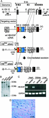
Generation of HB-EGF-null mice. (A) Gene-targeting strategy. Mouse HB-EGF cDNA containing the polyadenylation (pA) sequence flanked by loxP sequences was fused with the first exon of the mouse HB-EGF gene. The lacZ gene was inserted downstream of the HB-EGF cDNA. The targeting vector also contains the neomycin-resistance gene (neo), driven by the phosphoglycerate kinase (PGK) promoter, and the diphtheria toxin A-fragment (DT-A) gene. Cre-mediated recombination results in the deletion of HB-EGF cDNA and in the expression of the lacZ gene. Exon sequences are indicated as black boxes. cDNAs encoding HB-EGF and LacZ are indicated as red and blue boxes, respectively. The loxP sites are indicated by yellow-hatched triangles. E, _Eco_RI; H, _Hin_dIII; K, _Kpn_I; S, _Sac_II; V, _Eco_RV; X, _Xho_I. (B) Southern blot analysis of the recombination of the HBlox allele. Genomic DNA from tails of WT (+/+), HBlox/+ (lox/+), HBlox/lox (lox/lox), and HBdel/del (del/del) adult mice was digested with _Kpn_I and hybridized with a lox probe. This probe yields a 9.0-kb fragment from the WT allele, a 2.0-kb fragment from the HBlox allele, and a 5.3-kb fragment from the HBdel allele. (C) RT-PCR analysis of the expression of HB-EGF in WT (+/+) and HBdel/del (del/del) mice. Transcripts encoding HB-EGF were observed in the heart and kidney, but not in the liver, of WT mice. No HB-EGF transcripts were found in HBdel/del mice. GAPDH (G3PDH) mRNA was measured as a control for RNA preparation. (D) HB-EGF LacZ staining of a 12-wk-old HBdel/+ heart section. (Bar = 50 μm.)
Figure 2
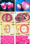
HB-EGF-null mice have heart defects. (A) Representative embryonic day 19.5 (E19.5) WT (+/+) and HBdel/del (del/del) hearts. (B) Representative 6-wk-old WT (+/+) and HBdel/del (del/del) hearts. (C_–_H) Hematoxylin/eosin staining of transverse sections of hearts at the papillary muscle level. (C and D) WT and HBdel/del hearts, respectively, from an E19.5 embryo. (E and F) WT and HBdel/del hearts, respectively, from 12-wk-old mice. In D blood clots are indicated by asterisks. (G and H) High-magnification pictures of E and F, respectively. (Bars = 2 mm in A and B, 300 μm in C and D, 3 mm in E and F, and 20 μm in G and H.)
Figure 3
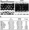
Echocardiography. (A) representative records of 2D echocardiograms of WT (+/+) and HBdel/del (del/del) mice. Arrows indicate the end-diastolic (d) and end-systolic (s) dimensions, respectively. (B) Physiological parameters of 8- to 12-wk-old mice. BW, body weight; HR, heart rate; sBP and dBP, systolic and diastolic blood pressure, respectively; FS, fractional shortening calculated as [(LVDd − LVDs)/LVDd] × 100; n.s., not significant. All values are ±SEM.
Figure 4
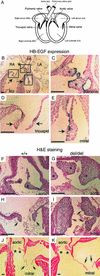
Cardiac valve defects in HBdel/del mice. (A) Schematic illustration of the heart depicting positions of the valves. (B_–_E) LacZ staining of longitudinal sections through the hearts of E19.5 HBdel/+ (corresponds to WT) embryos. (B) HB-EGF is expressed at the margins of the pulmonic valve (asterisks) and in tricuspid and mitral valves (arrows). pa, pulmonary artery. (C_–_E) Higher magnification of areas indicated by the boxes in B. Pulmonic valve (C, asterisks), tricuspid valve (D, arrows), and mitral valve (E, arrow) are shown. (F_–_K) Histological analysis of cardiac valves. Shown are hematoxylin/eosin-stained longitudinal sections of hearts of E19.5 embryos (F_–_I) and of 12-wk-old mice (J and K). Mice are WT (F, H, and J) and HBdel/del (G, I, and K). E19.5 aortic valves (F and G, asterisks), E19.5 mitral valves (H and I, arrows), and 12-wk-old aortic (J and K, asterisks) and mitral (J and K, arrows) valves are shown. Pulmonic and tricuspid valves are also enlarged (not shown). (Bars = 150 μm.)
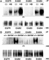
Figure 5
Tyrosine phosphorylation of the ErbB receptor family. (A) DMEM (−) or 5 μg/ml recombinant soluble HB-EGF (HB) was perfused directly into the WT heart. Each ErbB receptor was immunoprecipitated from lysates with its corresponding Ab. The immunoprecipitates (IP) were analyzed by Western blotting (WB) using anti-phosphotyrosine Ab (PY) or each of the four corresponding anti-ErbB Abs. (B) Comparison of the constitutive phosphorylation and protein levels of ErbB receptors in WT (+/+) and HBdel/del (del/del) hearts. Hearts perfused with DMEM were homogenized and analyzed by immunoprecipitation and Western blotting as in A.
Similar articles
- Defective valvulogenesis in HB-EGF and TACE-null mice is associated with aberrant BMP signaling.
Jackson LF, Qiu TH, Sunnarborg SW, Chang A, Zhang C, Patterson C, Lee DC. Jackson LF, et al. EMBO J. 2003 Jun 2;22(11):2704-16. doi: 10.1093/emboj/cdg264. EMBO J. 2003. PMID: 12773386 Free PMC article. - ErbB and HB-EGF signaling in heart development and function.
Iwamoto R, Mekada E. Iwamoto R, et al. Cell Struct Funct. 2006;31(1):1-14. doi: 10.1247/csf.31.1. Cell Struct Funct. 2006. PMID: 16508205 Review. - HB-EGF promotes epithelial cell migration in eyelid development.
Mine N, Iwamoto R, Mekada E. Mine N, et al. Development. 2005 Oct;132(19):4317-26. doi: 10.1242/dev.02030. Epub 2005 Sep 1. Development. 2005. PMID: 16141218 - Heparin-binding epidermal growth factor-like growth factor as a novel targeting molecule for cancer therapy.
Miyamoto S, Yagi H, Yotsumoto F, Kawarabayashi T, Mekada E. Miyamoto S, et al. Cancer Sci. 2006 May;97(5):341-7. doi: 10.1111/j.1349-7006.2006.00188.x. Cancer Sci. 2006. PMID: 16630129 Free PMC article. Review.
Cited by
- Heparin-binding EGF-like growth factor induces heart interstitial fibrosis via an Akt/mTor/p70s6k pathway.
Lian H, Ma Y, Feng J, Dong W, Yang Q, Lu D, Zhang L. Lian H, et al. PLoS One. 2012;7(9):e44946. doi: 10.1371/journal.pone.0044946. Epub 2012 Sep 12. PLoS One. 2012. PMID: 22984591 Free PMC article. - Lung cancer-derived galectin-1 enhances tumorigenic potentiation of tumor-associated dendritic cells by expressing heparin-binding EGF-like growth factor.
Kuo PL, Huang MS, Cheng DE, Hung JY, Yang CJ, Chou SH. Kuo PL, et al. J Biol Chem. 2012 Mar 23;287(13):9753-9764. doi: 10.1074/jbc.M111.321190. Epub 2012 Jan 30. J Biol Chem. 2012. PMID: 22291012 Free PMC article. - Hypoxia changes the expression of the epidermal growth factor (EGF) system in human hearts and cultured cardiomyocytes.
Munk M, Memon AA, Goetze JP, Nielsen LB, Nexo E, Sorensen BS. Munk M, et al. PLoS One. 2012;7(7):e40243. doi: 10.1371/journal.pone.0040243. Epub 2012 Jul 5. PLoS One. 2012. PMID: 22792252 Free PMC article. - Generation and characterization of conditional heparin-binding EGF-like growth factor knockout mice.
Oyagi A, Oida Y, Kakefuda K, Shimazawa M, Shioda N, Moriguchi S, Kitaichi K, Nanba D, Yamaguchi K, Furuta Y, Fukunaga K, Higashiyama S, Hara H. Oyagi A, et al. PLoS One. 2009 Oct 14;4(10):e7461. doi: 10.1371/journal.pone.0007461. PLoS One. 2009. PMID: 19829704 Free PMC article. - Mice with defects in HB-EGF ectodomain shedding show severe developmental abnormalities.
Yamazaki S, Iwamoto R, Saeki K, Asakura M, Takashima S, Yamazaki A, Kimura R, Mizushima H, Moribe H, Higashiyama S, Endoh M, Kaneda Y, Takagi S, Itami S, Takeda N, Yamada G, Mekada E. Yamazaki S, et al. J Cell Biol. 2003 Nov 10;163(3):469-75. doi: 10.1083/jcb.200307035. Epub 2003 Nov 3. J Cell Biol. 2003. PMID: 14597776 Free PMC article.
References
- Tzahar E, Yarden Y. Biochim Biophys Acta. 1998;1377:M25–M37. - PubMed
- Higashiyama S, Abraham J A, Miller J, Fiddes J C, Klagsbrun M A. Science. 1991;251:936–939. - PubMed
- Higashiyama S, Lau K, Besner G E, Abraham J A, Klagsbrun M. J Biol Chem. 1992;267:6205–6212. - PubMed
Publication types
MeSH terms
Substances
LinkOut - more resources
Full Text Sources
Other Literature Sources
Molecular Biology Databases
Research Materials
Miscellaneous
