Platelet-derived growth factor-B enhances glioma angiogenesis by stimulating vascular endothelial growth factor expression in tumor endothelia and by promoting pericyte recruitment - PubMed (original) (raw)
Platelet-derived growth factor-B enhances glioma angiogenesis by stimulating vascular endothelial growth factor expression in tumor endothelia and by promoting pericyte recruitment
Ping Guo et al. Am J Pathol. 2003 Apr.
Abstract
Platelet-derived growth factor (PDGF)-B and its receptor (PDGF-R) beta are overexpressed in human gliomas and responsible for recruiting peri-endothelial cells to vessels. To establish the role of PDGF-B in glioma angiogenesis, we overexpressed PDGF-B in U87MG glioma cells. Although PDGF-B stimulated tyrosine phosphorylation of PDGF-Rbeta in U87MG cells, treatment with recombinant PDGF-B or overexpression of PDGF-B in U87MG cells had no effect on their proliferation. However, an increase of secreted PDGF-B in conditioned media of U87MG/PDGF-B cells promoted migration of endothelial cells expressing PDGF-R beta, whereas conditioned media from U87MG cells did not increase the cell migration. In mice, overexpression of PDGF-B in U87MG cells enhanced intracranial glioma formation by stimulating vascular endothelial growth factor (VEGF) expression in neovessels and by attracting vessel-associated pericytes. When PDGF-B and VEGF were overexpressed simultaneously by U87MG tumors, there was a marked increase of capillary-associated pericytes as seen in U87MG/VEGF(165)/PDGF-B gliomas. As a result of pericyte recruitment, vessels induced by VEGF in tumor vicinity migrated into the central regions of these tumors. These data suggest that PDGF-B is a paracrine factor in U87MG gliomas, and that PDGF-B enhances glioma angiogenesis, at least in part, by stimulating VEGF expression in tumor endothelia and by recruiting pericytes to neovessels.
Figures
Figure 1.
PDGF-B activates endogenous PDGF-Rβ in U87MG glioma cells. PAE, PAE-PDGF-Rβ, and U87MG cells were serum-starved overnight. The cells were then treated with 50 ng/ml of a human recombinant PDGF-B protein at 37°C for 15 minutes. The cell lysates were subjected to immunoblotting for detection of tyrosine phosphorylation on PDGF-Rβ. The membrane was then reprobed with an anti-human PDGF-Rβ antibody. The PDGF-Rβ protein runs as a 180-kd band in a 7.5% SDS-PAGE gel (bottom). Duplicate experiments yielded identical results.
Figure 2.
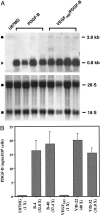
Expression of exogenous PDGF-B in U87MG glioma cells. A: Northern blot analysis. Top: Samples of the parental U87MG and two types of PDGF-B-overexpressing cells are shown. In each class, four individual PDGF-B-expressing clones are presented. Endogenous (•, 3.8 kb) and exogenous (▸, 0.8 kb) PDGF-B transcripts and their descriptions are in the text. Bottom: Methylene blue staining of the membrane after RNA was transferred into the membrane. ▪: 18 S and 28 S ribosomal RNA species. Duplicate experiments yielded similar results. B: PDGF-B ELISA analyses. Cells (3 × 10⁵) of the parental U87MG- or PDGF-B-expressing cells or VEGF165- or VEGF165/PDGF-B-expressing cells were seeded onto 12-well plates in triplicate. On the next day, the media was replaced with DMEM/0.5% bovine serum albumin/1% dialyzed fetal bovine serum for another 24 hours. CM was collected after the cells were cultured for an additional 48 hours. Each bar represents the mean ± SEM of three triplicates. Identical experiments were also performed using CM of other PDGF-B-overexpressing clones, B-26, B-34, V/B-11, and V/B-33. The assays were performed two additional times using cells of various passage numbers with similar results.
Figure 3.
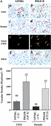
PDGF-B enhances U87MG glioma angiogenesis by increasing VEGF expression in tumor ECs and by recruiting vessel-associated pericytes. A: Immunohistochemical analyses of gliomas formed by the parental U87MG- and PDGF-B-expressing cells. The analyses were performed with a rat monoclonal anti-CD31 antibody (a and b), a polyclonal anti-desmin antibody (c and d), a polyclonal anti-VEGF antibody (red color) together with the anti-CD31 antibody (green color, e and f) and a polyclonal anti-PDGF-B antibody (g and h). Arrows show positive staining for blood vessels (a and b) or pericytes (c and d). In e and f, arrows indicate blood vessels in the tumors. Arrowheads indicate VEGF staining. In f, most of the vessels were stained by both the anti-VEGF and the anti-CD31 (vessels) antibodies (orange color). a, c, b, and d: Immunohistochemical staining in identical areasof serial sections from the same individual mouse brains. Six or more individual tumor samples of each type were analyzed. Experiments were repeated two additional times with similar results. B: Increased vessel densities and recruitment of vessel-associated pericytes in U87MG PDGF-B-expressing gliomas. Quantitative analyses of immunohistochemistry data that are shown in a, b, c, and d were done as described in Materials and Methods. The representative immunohistochemical stains from the parental U87MG- and PDGF-B-expressing gliomas are shown in a and b (vessel stains) or c and d (desmin stains). In each analysis, 10 to 15 random areas within the same tissue section were examined. The mean values of five to seven serial sections from six to seven separate mouse brains in each group were used for the quantitative analyses. Data are presented as means ± SD. Numbers above each column are the numbers of mice analyzed in each group. Numbers in the parentheses under the x axis are the fold difference of the densities found in U87MG- and PDGF-B-expressing gliomas compared with that in parental U87MG tumors. Original magnifications in A: ×200 (a, b); ×400 (c, d, e, f, g, and h).
Figure 4.
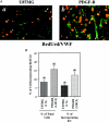
Overexpression of PDGF-B increased both tumor and EC proliferation. A: Immunohistochemical analyses of gliomas formed by parental U87MG- and PDGF-B-expressing cells. The analyses were performed with a monoclonal anti-BrdUrd antibody together with a polyclonal anti-von Willebrand factor antibody (a and b). Arrows show positive staining of proliferative nuclei in blood vessels. Arrowheads indicate BrdUrd staining only in the nuclei of tumor cells. Three to five serial sections from four to six individual tumor samples of each type were analyzed independently. Experiments were repeated two additional times with similar results. B: Increased proliferative index in both total cell (left columns) and ECs (right columns) in the PDGF-B-expressing gliomas. Quantitative analyses of immunohistochemistry data were done as described in Materials and Methods. Original magnification: ×400 (A).
Figure 5.
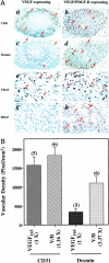
PDGF-B augments U87MG glioma angiogenesis by promoting vessel migration and recruiting vessel-associated pericytes. A: Immunohistochemical analyses of gliomas formed by either VEGF165- or VEGF165/PDGF-B-expressing cells. The analyses were performed with the anti-CD31 antibody (a and b), the polyclonal anti-desmin antibody (c and d), the polyclonal anti-VEGF antibody (e and f), and the polyclonal anti-PDGF-B antibody (g and h). Arrows show positive staining for blood vessels (a and b) or pericytes (c and d) or VEGF (e and f) or PDGF-B (g and h). Areas shown in a, c, and b, d or e, g, and f, h are immunohistochemical staining in identical areas from the same individual mouse brains. Five serial sections from five or six individual tumor samples of each type were analyzed independently. Experiments were repeated two independent times with similar results. B: Increased recruitment of vessel-associated pericytes in U87MG/VEGF/PDGF-B-expressing gliomas. Quantitative analyses of immunohistochemistry data were performed as described in Materials and Methods. Original magnifications in A: ×40 (a, b, c, and d); ×400 (e, f, g, and h).
Similar articles
- A role for VEGF as a negative regulator of pericyte function and vessel maturation.
Greenberg JI, Shields DJ, Barillas SG, Acevedo LM, Murphy E, Huang J, Scheppke L, Stockmann C, Johnson RS, Angle N, Cheresh DA. Greenberg JI, et al. Nature. 2008 Dec 11;456(7223):809-13. doi: 10.1038/nature07424. Epub 2008 Nov 9. Nature. 2008. PMID: 18997771 Free PMC article. - Tumor-derived vascular endothelial growth factor up-regulates angiopoietin-2 in host endothelium and destabilizes host vasculature, supporting angiogenesis in ovarian cancer.
Zhang L, Yang N, Park JW, Katsaros D, Fracchioli S, Cao G, O'Brien-Jenkins A, Randall TC, Rubin SC, Coukos G. Zhang L, et al. Cancer Res. 2003 Jun 15;63(12):3403-12. Cancer Res. 2003. PMID: 12810677 - Vascular endothelial growth factor acts as a pericyte mitogen under hypoxic conditions.
Yamagishi S, Yonekura H, Yamamoto Y, Fujimori H, Sakurai S, Tanaka N, Yamamoto H. Yamagishi S, et al. Lab Invest. 1999 Apr;79(4):501-9. Lab Invest. 1999. PMID: 10212003 - Possible involvement of VEGF-FLT tyrosine kinase receptor system in normal and tumor angiogenesis.
Shibuya M, Seetharam L, Ishii Y, Sawano A, Gotoh N, Matsushime H, Yamaguchi S. Shibuya M, et al. Princess Takamatsu Symp. 1994;24:162-70. Princess Takamatsu Symp. 1994. PMID: 8983073 Review.
Cited by
- Molecular mechanisms of the effect of TGF-β1 on U87 human glioblastoma cells.
Bryukhovetskiy I, Shevchenko V. Bryukhovetskiy I, et al. Oncol Lett. 2016 Aug;12(2):1581-1590. doi: 10.3892/ol.2016.4756. Epub 2016 Jun 22. Oncol Lett. 2016. PMID: 27446475 Free PMC article. - Lessons from anti-vascular endothelial growth factor and anti-vascular endothelial growth factor receptor trials in patients with glioblastoma.
Lu-Emerson C, Duda DG, Emblem KE, Taylor JW, Gerstner ER, Loeffler JS, Batchelor TT, Jain RK. Lu-Emerson C, et al. J Clin Oncol. 2015 Apr 1;33(10):1197-213. doi: 10.1200/JCO.2014.55.9575. Epub 2015 Feb 23. J Clin Oncol. 2015. PMID: 25713439 Free PMC article. - Endothelial-Tumor Cell Interaction in Brain and CNS Malignancies.
Peleli M, Moustakas A, Papapetropoulos A. Peleli M, et al. Int J Mol Sci. 2020 Oct 6;21(19):7371. doi: 10.3390/ijms21197371. Int J Mol Sci. 2020. PMID: 33036204 Free PMC article. Review. - Combined effects of pericytes in the tumor microenvironment.
Ribeiro AL, Okamoto OK. Ribeiro AL, et al. Stem Cells Int. 2015;2015:868475. doi: 10.1155/2015/868475. Epub 2015 Apr 27. Stem Cells Int. 2015. PMID: 26000022 Free PMC article. Review. - Glial progenitors in adult white matter are driven to form malignant gliomas by platelet-derived growth factor-expressing retroviruses.
Assanah M, Lochhead R, Ogden A, Bruce J, Goldman J, Canoll P. Assanah M, et al. J Neurosci. 2006 Jun 21;26(25):6781-90. doi: 10.1523/JNEUROSCI.0514-06.2006. J Neurosci. 2006. PMID: 16793885 Free PMC article.
References
- Carmeliet P: Mechanisms of angiogenesis and arteriogenesis. Nat Med 2000, 6:389-395 - PubMed
- Ferrara N: Vascular endothelial growth factor: molecular and biological aspects. Curr Top Microbiol Immunol 1999, 237:1-30 - PubMed
- Yancopoulos GD, Davis S, Gale NW, Rudge JS, Wiegand SJ, Holash J: Vascular-specific growth factors and blood vessel formation. Nature 2000, 407:242-248 - PubMed
- Hirschi KK, Rohovsky SA, D’Amore PA: PDGF, TGF-beta, and heterotypic cell-cell interactions mediate endothelial cell-induced recruitment of 10T1/2 cells and their differentiation to a smooth muscle fate [published erratum appears in J Cell Biol 1998 Jun 1;141(5):1287]. J Cell Biol 1998, 141:805-814 - PMC - PubMed
- Hellstrom M, Kalen M, Lindahl P, Abramsson A, Betsholtz C: Role of PDGF-B and PDGFR-beta in recruitment of vascular smooth muscle cells and pericytes during embryonic blood vessel formation in the mouse. Development 1999, 126:3047-3055 - PubMed
Publication types
MeSH terms
Substances
LinkOut - more resources
Full Text Sources
Other Literature Sources
Research Materials
Miscellaneous


