Molecular paleontology of transposable elements in the Drosophila melanogaster genome - PubMed (original) (raw)
Molecular paleontology of transposable elements in the Drosophila melanogaster genome
Vladimir V Kapitonov et al. Proc Natl Acad Sci U S A. 2003.
Abstract
We report here a superfamily of "cut and paste" DNA transposons called Transib. These transposons populate the Drosophila melanogaster and Anopheles gambiae genomes, use a transposase that is not similar to any known proteins, and are characterized by 5-bp target site duplications. We found that the fly genome, which was thought to be colonized by the P element <100 years ago, harbors approximately 5 million year (Myr)-old fossils of ProtoP, an ancient ancestor of the P element. We also show that Hoppel, a previously reported transposable element (TE), is a nonautonomous derivate of ProtoP. We found that the "rolling-circle" Helitron transposons identified previously in plants and worms populate also insect genomes. Our results indicate that Helitrons were horizontally transferred into the fly or/and mosquito genomes. We have also identified a most abundant TE in the fly genome, DNAREP1_DM, which is an approximately 10-Myr-old footprint of a Penelope-like retrotransposon. We estimated that TEs are three times more abundant than reported previously, making up approximately 22% of the whole genome. The chromosomal and age distributions of TEs in D. melanogaster are very similar to those in Arabidopsis thaliana. Both genomes contain only relatively young TEs (<20 Myr old), constituting a main component of paracentromeric regions.
Figures
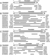
Fig. 1.
Reconstruction of the Transib1 (A), Transib2 (B), Transib3 (C), and Transib4 (D) consensus sequences. The consensus sequences are schematically depicted as rectangles capped by black triangles indicating TIRs. Transposasecoding regions are shaded in gray. Their coordinates in the consensus sequences are shown above the rectangles. Transib copies used for reconstruction of the consensus sequences are shown as thick lines beneath the rectangles. Gaps in the lines mark deletions. GenBank accession numbers and the corresponding sequence coordinates of TEs are indicated.
Fig. 2.
Multiple alignment of the Transib transposases. Diamonds mark amino acid residues that belong to a putative noncanonical DDE catalytic site. Asterisks mark bp 10, 30, etc. Conserved motifs and residues are underlined and shaded, respectively.
Fig. 3.
Reconstruction of the ProtoP consensus sequence. The consensus sequence is schematically depicted as a rectangle capped by the black triangles indicating TIRs. The region coding for the transposase is shaded in gray. Its coordinates in the consensus sequence are indicated above the rectangle. Copies of ProtoP used for reconstructing the consensus sequences are shown as thick lines beneath the ProtoP, Hoppel, and ProtoP_ B consensus sequences. Gaps in the lines mark deletions. GenBank accession numbers and the corresponding sequence coordinates of TEs are indicated.
Fig. 4.
A phylogenetic tree for _P_-like transposases. The unrooted tree was constructed by using the neighbor-joining method implemented in MEGA (19). Transposases from the next _P_-like transposons are shown: P_ DH, Drosophila helvetica, GenBank accession no. AAK08181; P_ SP, Scaptomyza pallida, joined AAA29959–61; P_ DM, DM, A24786; P_ DB, Drosophila bifasciata, AAB31526; P_ LC, Lucilia cuprina, A46361. P1_ AG and P3_ AG from A. gambiae and ProtoP from DM are transposases from P elements reported in this manuscript. HS is a putative human gene derived from the _P_-like transposase (27). The scale of the Poisson correction distances between the protein sequences is indicated. Bootstrap values are shown at the nodes.
Fig. 5.
Density of TEs across chromosome 2. It was calculated as a percentage of TE-derived sequences per 100 kb in nonoverlapping windows. The ≈8-megabase (Mb) centromeric region is shaded in gray.
Similar articles
- Species-specific chromatin landscape determines how transposable elements shape genome evolution.
Huang Y, Shukla H, Lee YCG. Huang Y, et al. Elife. 2022 Aug 23;11:e81567. doi: 10.7554/eLife.81567. Elife. 2022. PMID: 35997258 Free PMC article. - Molecular paleontology of transposable elements from Arabidopsis thaliana.
Kapitonov VV, Jurka J. Kapitonov VV, et al. Genetica. 1999;107(1-3):27-37. Genetica. 1999. PMID: 10952195 - Rolling-circle transposons in eukaryotes.
Kapitonov VV, Jurka J. Kapitonov VV, et al. Proc Natl Acad Sci U S A. 2001 Jul 17;98(15):8714-9. doi: 10.1073/pnas.151269298. Epub 2001 Jul 10. Proc Natl Acad Sci U S A. 2001. PMID: 11447285 Free PMC article. - Helitrons on a roll: eukaryotic rolling-circle transposons.
Kapitonov VV, Jurka J. Kapitonov VV, et al. Trends Genet. 2007 Oct;23(10):521-9. doi: 10.1016/j.tig.2007.08.004. Epub 2007 Sep 11. Trends Genet. 2007. PMID: 17850916 Review. - Constitutive heterochromatin and transposable elements in Drosophila melanogaster.
Dimitri P. Dimitri P. Genetica. 1997;100(1-3):85-93. Genetica. 1997. PMID: 9440261 Review.
Cited by
- Regulatory Changes in the Fatty Acid Elongase eloF Underlie the Evolution of Sex-specific Pheromone Profiles in Drosophila prolongata.
Luo Y, Takau A, Li J, Fan T, Hopkins BR, Le Y, Ramirez SR, Matsuo T, Kopp A. Luo Y, et al. bioRxiv [Preprint]. 2024 Oct 14:2024.10.09.617394. doi: 10.1101/2024.10.09.617394. bioRxiv. 2024. PMID: 39464098 Free PMC article. Preprint. - Comprehensive analysis of the Xya riparia genome uncovers the dominance of DNA transposons, LTR/Gypsy elements, and their evolutionary dynamics.
Khan H, Yuan H, Liu X, Nie Y, Majid M. Khan H, et al. BMC Genomics. 2024 Jul 12;25(1):687. doi: 10.1186/s12864-024-10596-5. BMC Genomics. 2024. PMID: 38997681 Free PMC article. - Genomes of historical specimens reveal multiple invasions of LTR retrotransposons in Drosophila melanogaster during the 19th century.
Scarpa A, Pianezza R, Wierzbicki F, Kofler R. Scarpa A, et al. Proc Natl Acad Sci U S A. 2024 Apr 9;121(15):e2313866121. doi: 10.1073/pnas.2313866121. Epub 2024 Apr 2. Proc Natl Acad Sci U S A. 2024. PMID: 38564639 Free PMC article. - Spoink, a LTR retrotransposon, invaded D. melanogaster populations in the 1990s.
Pianezza R, Scarpa A, Narayanan P, Signor S, Kofler R. Pianezza R, et al. PLoS Genet. 2024 Mar 26;20(3):e1011201. doi: 10.1371/journal.pgen.1011201. eCollection 2024 Mar. PLoS Genet. 2024. PMID: 38530818 Free PMC article. - ChimeraTE: a pipeline to detect chimeric transcripts derived from genes and transposable elements.
Oliveira DS, Fablet M, Larue A, Vallier A, Carareto CMA, Rebollo R, Vieira C. Oliveira DS, et al. Nucleic Acids Res. 2023 Oct 13;51(18):9764-9784. doi: 10.1093/nar/gkad671. Nucleic Acids Res. 2023. PMID: 37615575 Free PMC article.
References
- Berg, D. E. & Howe, M. H., eds. (1987) Mobile DNA (Am. Soc. Microbiol., Washington, DC).
- Kidwell, M. G. & Lisch, D. R. (2001) Evol. Int. J. Org. Evol. 55, 1–24. - PubMed
- Fedoroff, N. V. (1999) Ann. N.Y. Acad. Sci. 870, 251–264. - PubMed
- Craig, N. L. (1995) Science 270, 253–254. - PubMed
- Adams, M. D., Celniker, S. E., Holt, R. A., Evans, C. A., Gocayne, J. D., Amanatides, P. G., Scherer, S. E., Li, P. W., Hoskins, R. A., Galle, R. F., et al. (2000) Science 287, 2185–2195. - PubMed
Publication types
MeSH terms
Substances
LinkOut - more resources
Full Text Sources
Molecular Biology Databases


