An EP2 receptor-selective prostaglandin E2 agonist induces bone healing - PubMed (original) (raw)
. 2003 May 27;100(11):6736-40.
doi: 10.1073/pnas.1037343100. Epub 2003 May 14.
F Borovecki, H Z Ke, K O Cameron, B Lefker, W A Grasser, T A Owen, M Li, P DaSilva-Jardine, M Zhou, R L Dunn, F Dumont, R Korsmeyer, P Krasney, T A Brown, D Plowchalk, S Vukicevic, D D Thompson
Affiliations
- PMID: 12748385
- PMCID: PMC164516
- DOI: 10.1073/pnas.1037343100
An EP2 receptor-selective prostaglandin E2 agonist induces bone healing
V M Paralkar et al. Proc Natl Acad Sci U S A. 2003.
Abstract
The morbidity and mortality associated with impaired/delayed fracture healing remain high. Our objective was to identify a small nonpeptidyl molecule with the ability to promote fracture healing and prevent malunions. Prostaglandin E2 (PGE2) causes significant increases in bone mass and bone strength when administered systemically or locally to the skeleton. However, due to side effects, PGE2 is an unacceptable therapeutic option for fracture healing. PGE2 mediates its tissue-specific pharmacological activity via four different G protein-coupled receptor subtypes, EP1, -2, -3, and -4. The anabolic action of PGE2 in bone has been linked to an elevated level of cAMP, thereby implicating the EP2 and/or EP4 receptor subtypes in bone formation. We identified an EP2 selective agonist, CP-533,536, which has the ability to heal canine long bone segmental and fracture model defects without the objectionable side effects of PGE2, suggesting that the EP2 receptor subtype is a major contributor to PGE2's local bone anabolic activity. The potent bone anabolic activity of CP-533,536 offers a therapeutic alternative for the treatment of fractures and bone defects in patients.
Figures
Fig. 1.
(A) Chemical structure of CP-533,536. (B) Analysis of intracellular cAMP levels on treatment of cells with CP-533,536. HEK-293 cells stably transfected with the EP2 receptor were treated with increasing concentrations of CP-533,536, and intracellular cAMP levels were measured.
Fig. 2.
Peripheral quantitative computerized tomography images of proximal tibial cross sections from rats injected with vehicle or CP-533,536 directly into the bone marrow. White area, within the bone circumference, represents areas of high bone mass, whereas other colors represent areas of lower-density tissues including bone marrow (red).
Fig. 3.
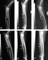
X-rays of canine ulnar critical defect treated with 1.0 ml of PLGH matrix show no healing/rebridging sequence at 2 (A), 12 (B), and 24 (C) weeks after surgery. Critical defects treated with 10 mg of CP-533,536 dissolved in 1.0 ml of matrix showed a time-dependent healing/rebridging sequence at 2 (D), 12 (E), and 24 (F) weeks after surgery.
Fig. 4.
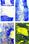
A toluidine blue-stained section of the midulnar region from the canine critical defect model after treatment with 1.0 ml of PLGH matrix alone shows no rebridgement at 24 weeks after surgery (A), whereas full rebridgement was observed after treatment with 10 mg of CP-533,536 (B). Intense remodeling of the newly formed cortical bone was observed and consisted of osteons (C, arrow-head), active vascular channels with hematopoietic marrow, rows of osteoblasts, and newly deposited osteoid (O) on the surface of mineralized lamellar bone (D, arrow). Final magnification: A and B = ×25; C and D = ×125.
Fig. 5.
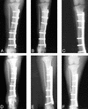
X-rays of a canine tibial osteotomy treated with 0.5 ml of PLGH matrix alone show no healing/rebridging sequence at 2 (A), 4 (B), and 8 (C) weeks after surgery. Defects treated with 5 mg of CP-533,536 dissolved in 0.5 ml of matrix showed healing/rebridging sequence at 2 (D), 4 (E), and 8 (F) weeks after surgery.
Fig. 6.
Plasma concentrations of CP-533,536 in dogs after administration in a PLGH matrix formulation to the site of a tibial osteotomy (•, 25 mg) or ulnar critical defect [▴, 10mg;♦,50mg;▪, 10 mg (0.2 ml of 50 mg/ml)]. Each point represents drug blood values from 4–10 dogs.
Similar articles
- A novel, non-prostanoid EP2 receptor-selective prostaglandin E2 agonist stimulates local bone formation and enhances fracture healing.
Li M, Ke HZ, Qi H, Healy DR, Li Y, Crawford DT, Paralkar VM, Owen TA, Cameron KO, Lefker BA, Brown TA, Thompson DD. Li M, et al. J Bone Miner Res. 2003 Nov;18(11):2033-42. doi: 10.1359/jbmr.2003.18.11.2033. J Bone Miner Res. 2003. PMID: 14606517 - Prostaglandin EP2 and EP4 receptor agonists in bone formation and bone healing: In vivo and in vitro evidence.
Graham S, Gamie Z, Polyzois I, Narvani AA, Tzafetta K, Tsiridis E, Helioti M, Mantalaris A, Tsiridis E. Graham S, et al. Expert Opin Investig Drugs. 2009 Jun;18(6):746-66. doi: 10.1517/13543780902893051. Expert Opin Investig Drugs. 2009. PMID: 19426119 Review. - Discovery of CP-533536: an EP2 receptor selective prostaglandin E2 (PGE2) agonist that induces local bone formation.
Cameron KO, Lefker BA, Ke HZ, Li M, Zawistoski MP, Tjoa CM, Wright AS, DeNinno SL, Paralkar VM, Owen TA, Yu L, Thompson DD. Cameron KO, et al. Bioorg Med Chem Lett. 2009 Apr 1;19(7):2075-8. doi: 10.1016/j.bmcl.2009.01.059. Epub 2009 Jan 23. Bioorg Med Chem Lett. 2009. PMID: 19250823 - Prostaglandin E2 activates EP2 receptors to inhibit human lung mast cell degranulation.
Kay LJ, Yeo WW, Peachell PT. Kay LJ, et al. Br J Pharmacol. 2006 Apr;147(7):707-13. doi: 10.1038/sj.bjp.0706664. Br J Pharmacol. 2006. PMID: 16432506 Free PMC article. - Prostaglandin E(2) receptors in bone formation.
Li M, Thompson DD, Paralkar VM. Li M, et al. Int Orthop. 2007 Dec;31(6):767-72. doi: 10.1007/s00264-007-0406-x. Epub 2007 Jun 26. Int Orthop. 2007. PMID: 17593365 Free PMC article. Review.
Cited by
- Influence of Pain and Analgesia on Orthopedic and Wound-healing Models in Rats and Mice.
Huss MK, Felt SA, Pacharinsak C. Huss MK, et al. Comp Med. 2019 Dec 1;69(6):535-545. doi: 10.30802/AALAS-CM-19-000013. Epub 2019 Sep 27. Comp Med. 2019. PMID: 31561753 Free PMC article. Review. - Wound Healing Versus Regeneration: Role of the Tissue Environment in Regenerative Medicine.
Atala A, Irvine DJ, Moses M, Shaunak S. Atala A, et al. MRS Bull. 2010 Aug 1;35(8):10.1557/mrs2010.528. doi: 10.1557/mrs2010.528. MRS Bull. 2010. PMID: 24241586 Free PMC article. - Recombinant Human Bone Morphogenetic Protein 6 Delivered Within Autologous Blood Coagulum Restores Critical Size Segmental Defects of Ulna in Rabbits.
Grgurevic L, Oppermann H, Pecin M, Erjavec I, Capak H, Pauk M, Karlovic S, Kufner V, Lipar M, Bubic Spoljar J, Bordukalo-Niksic T, Maticic D, Peric M, Windhager R, Sampath TK, Vukicevic S. Grgurevic L, et al. JBMR Plus. 2018 Nov 5;3(5):e10085. doi: 10.1002/jbm4.10085. eCollection 2019 May. JBMR Plus. 2018. PMID: 31131338 Free PMC article. - Impact of Bone Morphogenetic Protein 7 and Prostaglandin receptors on osteoblast healing and organization of collagen.
Salama MA, Anwar Ismail A, Islam MS, K G AR, Al Kawas S, Samsudin AR, A C SA. Salama MA, et al. PLoS One. 2024 May 16;19(5):e0303202. doi: 10.1371/journal.pone.0303202. eCollection 2024. PLoS One. 2024. PMID: 38753641 Free PMC article. - Altered bone biology in psoriatic arthritis.
Rahimi H, Ritchlin CT. Rahimi H, et al. Curr Rheumatol Rep. 2012 Aug;14(4):349-57. doi: 10.1007/s11926-012-0259-1. Curr Rheumatol Rep. 2012. PMID: 22592745 Free PMC article. Review.
References
- Lieberman, J. R., Daluiski, A. & Einhorn, T. A. (2002) J. Bone Joint Surg. 84-A, 1032-1044. - PubMed
- Bostrom, M. P. G., Yang, X. & Koutras, I. (2000) Curr. Opin. Orthop. 11, 403-412.
- Barnes, G. L., Kostenuik, P. J., Gerstenfeld, L. C. & Einhorn, T. A. (1999) J. Bone Miner. Res. 14, 1805-1815. - PubMed
- Einhorn, T. A. (1995) J. Bone Joint Surg. 77-A, 940-956. - PubMed
- Rueger, D. C. (2002) in Bone Morphogenetic Proteins: From Laboratory to Clinical Practice, eds. Vukicevic, S. & Sampath, K. T. (Birkhauser, Basel), pp. 1-19.
MeSH terms
Substances
LinkOut - more resources
Full Text Sources
Other Literature Sources
Molecular Biology Databases
Miscellaneous


