Transient activation of beta -catenin signaling in cutaneous keratinocytes is sufficient to trigger the active growth phase of the hair cycle in mice - PubMed (original) (raw)
Transient activation of beta -catenin signaling in cutaneous keratinocytes is sufficient to trigger the active growth phase of the hair cycle in mice
David Van Mater et al. Genes Dev. 2003.
Abstract
Wnts have key roles in many developmental processes, including hair follicle growth and differentiation. Stabilization of beta-catenin is essential in the canonical Wnt signaling pathway. We developed transgenic mice expressing a regulated form of beta-catenin in the skin. Chronic activation of beta-catenin in resting (telogen) hair follicles resulted in changes consistent with induction of an exaggerated, aberrant growth phase (anagen). Transient activation of beta-catenin produced a normal anagen. Our data lend strong support to the notion that a Wnt/beta-catenin signal operating on hair follicle precursor cells serves as a crucial proximal signal for the telogen-anagen transition.
Figures
Figure 1
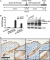
Activation of the K5/S33Yβ-catenin–ER protein by 4-OHT and expression in transgenic mice. (A) Schematic of the K5/S33Yβ-catenin–ER construct. A cDNA encoding the chimeric protein was subcloned into a bovine K5 expression cassette. (B) Activity of the K5/S33Yβ-catenin–ER protein is inducible by 4-OHT in 1811 keratinocytes. The 1811 keratinocytes were transfected with an empty K5 cassette, the pBabe/S33Yβ-catenin–ER construct, or the K5/S33Yβ-catenin–ER construct along with either the TCF-responsive reporter construct TOPFLASH or control FOPFLASH construct. Cells were then treated with either 4-OHT in ethanol, or ethanol alone, and harvested 30 h later to assess luciferase activity. The assays were performed in duplicate; data are reported as the ratio of relative light units for TOPFLASH:FOPFLASH, normalized for transfection efficiency. (C) Expression of the K5/S33Yβ-catenin–ER fusion protein relative to endogenous β-catenin in transgenic mouse lines. Protein was isolated from tail skin of F1 mice derived from three independently derived founders and subjected to Western blot analysis with a mouse monoclonal anti-β-catenin antibody. The blot was reprobed with an anti-β-actin antibody to verify equal loading and transfer. (D) β-catenin protein localization in a clipped region of dorsal skin from K5/S33Yβ-catenin–ER transgenic mice not treated (−4-OHT) or 24 h after treatment with a single topical dose of 4-OHT (+4-OHT).
Figure 2
Growth and differentiation of the hair follicle in K5/S33Yβ-catenin–ER mice treated daily with 4-OHT. A region of dorsal hair was clipped on both transgenic and wild-type littermates and the skin area was treated daily with 4-OHT in ethanol. Parasagittal sections of skin were taken at time points following initiation of treatment and stained with hematoxylin and eosin. Hair follicles in wild-type mice remained in the resting phase (telogen) throughout the experiment; growth and differentiation of transgenic hair follicles was dramatically stimulated.
Figure 3
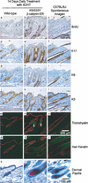
Altered expression of hair follicle markers in K5/S33Yβ-catenin–ER mice. Immunohistochemistry and immunofluorescence were performed on parasagittal sections of skin from both wild-type and K5/S33Yβ-catenin–ER mice in addition to a normal, spontaneous anagen cycle in a C57BL/6J mouse. The markers used were as follows: BrdU (a–c), K17 (d–f), K6 (g–i), K5 (j–l), trichohyalin (m–o), hair keratin (p–r), and alkaline phosphatase staining for dermal papillae (s–u). White arrows highlight ectopic trichohyalin staining in n. Bars: a–l,s–u, 100 μm; m–r, 200 μm.
Figure 4
Transient treatment with 4-OHT is sufficient for normal anagen induction in K5/S33Yβ-catenin–ER mice. (A) Hair growth in K5/S33Yβ-catenin–ER mice following a single topical treatment with 4-OHT. The experiment was performed on L2 transgenic mice obtained following backcrossing to C57BL/6J mice for six generations. Hair on wild-type and transgenic littermates was clipped and the skin region treated with a single dose of 4-OHT in ethanol or ethanol alone. Hair growth at 17 d after 4-OHT treatment is shown. (B) Histology of hair follicles in K5/S33Yβ-catenin–ER mice over time after a single treatment with 4-OHT. A cohort of transgenic mice was clipped and treated with 4-OHT and followed for a period of 28 d. Parasagittal skin sections obtained at various times after 4-OHT treatment were stained with hematoxylin and eosin. Bar, 100 μm.
Similar articles
- Beta-catenin and Hedgehog signal strength can specify number and location of hair follicles in adult epidermis without recruitment of bulge stem cells.
Silva-Vargas V, Lo Celso C, Giangreco A, Ofstad T, Prowse DM, Braun KM, Watt FM. Silva-Vargas V, et al. Dev Cell. 2005 Jul;9(1):121-31. doi: 10.1016/j.devcel.2005.04.013. Dev Cell. 2005. PMID: 15992546 - [Regulation of hair follicle cycle].
Goryachkina VL, Ivanova MY, Tsomartova DA, Kartashkina NL, Kuznetsov SL. Goryachkina VL, et al. Morfologiia. 2014;146(5):83-7. Morfologiia. 2014. PMID: 25823297 Review. Russian. - STAT5 Activation in the Dermal Papilla Is Important for Hair Follicle Growth Phase Induction.
Legrand JMD, Roy E, Ellis JJ, Francois M, Brooks AJ, Khosrotehrani K. Legrand JMD, et al. J Invest Dermatol. 2016 Sep;136(9):1781-1791. doi: 10.1016/j.jid.2016.04.014. Epub 2016 Apr 27. J Invest Dermatol. 2016. PMID: 27131881 Review.
Cited by
- Effects of UV Induced-Photoaging on the Hair Follicle Cycle of C57BL6/J Mice.
Zhai X, Gong M, Peng Y, Yang D. Zhai X, et al. Clin Cosmet Investig Dermatol. 2021 May 18;14:527-539. doi: 10.2147/CCID.S310487. eCollection 2021. Clin Cosmet Investig Dermatol. 2021. PMID: 34040410 Free PMC article. - A Mouse Model for Conditional Expression of Activated β-Catenin in Epidermal Keratinocytes.
Maurya VK, Ying Y, Lydon JP. Maurya VK, et al. Transgenic Res. 2024 Oct;33(5):513-525. doi: 10.1007/s11248-024-00402-z. Epub 2024 Aug 7. Transgenic Res. 2024. PMID: 39110314 Free PMC article. - Home sweet home: skin stem cell niches.
Goldstein J, Horsley V. Goldstein J, et al. Cell Mol Life Sci. 2012 Aug;69(15):2573-82. doi: 10.1007/s00018-012-0943-3. Epub 2012 Mar 13. Cell Mol Life Sci. 2012. PMID: 22410738 Free PMC article. Review. - The evolving roles of Wnt signaling in stem cell proliferation and differentiation, the development of human diseases, and therapeutic opportunities.
Yu M, Qin K, Fan J, Zhao G, Zhao P, Zeng W, Chen C, Wang A, Wang Y, Zhong J, Zhu Y, Wagstaff W, Haydon RC, Luu HH, Ho S, Lee MJ, Strelzow J, Reid RR, He TC. Yu M, et al. Genes Dis. 2023 Jul 22;11(3):101026. doi: 10.1016/j.gendis.2023.04.042. eCollection 2024 May. Genes Dis. 2023. PMID: 38292186 Free PMC article. Review. - Modelling hair follicle growth dynamics as an excitable medium.
Murray PJ, Maini PK, Plikus MV, Chuong CM, Baker RE. Murray PJ, et al. PLoS Comput Biol. 2012;8(12):e1002804. doi: 10.1371/journal.pcbi.1002804. Epub 2012 Dec 20. PLoS Comput Biol. 2012. PMID: 23284275 Free PMC article.
References
- Andl T, Reddy ST, Gaddapara T, Millar SE. WNT signals are required for the initiation of hair follicle development. Dev Cell. 2002;2:643–653. - PubMed
- Chanda S, Robinette CL, Couse JF, Smart RC. 17β-estradiol and ICI-182780 regulate the hair follicle cycle in mice through an estrogen receptor-α pathway. Am J Physiol Endocrinol Metab. 2000;278:E202–E210. - PubMed
- DasGupta R, Fuchs E. Multiple roles for activated LEF/TCF transcription complexes during hair follicle development and differentiation. Development. 1999;126:4557–4568. - PubMed
- Fuchs E, Merrill BJ, Jamora C, DasGupta R. At the roots of a never-ending cycle. Dev Cell. 2001;1:13–25. - PubMed
- Gat U, DasGupta R, Degenstein L, Fuchs E. De Novo hair follicle morphogenesis and hair tumors in mice expressing a truncated beta-catenin in skin. Cell. 1998;95:605–614. - PubMed
Publication types
MeSH terms
Substances
Grants and funding
- T32GM07863/GM/NIGMS NIH HHS/United States
- P30 CA046592/CA/NCI NIH HHS/United States
- R01 CA085463/CA/NCI NIH HHS/United States
- T32 GM007863/GM/NIGMS NIH HHS/United States
- CA46592/CA/NCI NIH HHS/United States
- R01 AR045973/AR/NIAMS NIH HHS/United States
- R01 CA087837/CA/NCI NIH HHS/United States
- CA85463/CA/NCI NIH HHS/United States
- CA87837/CA/NCI NIH HHS/United States
LinkOut - more resources
Full Text Sources
Other Literature Sources
Molecular Biology Databases


