Structural variations and stabilising modifications of synthetic siRNAs in mammalian cells - PubMed (original) (raw)
Structural variations and stabilising modifications of synthetic siRNAs in mammalian cells
Frank Czauderna et al. Nucleic Acids Res. 2003.
Abstract
Double-stranded short interfering RNAs (siRNA) induce post-transcriptional silencing in a variety of biological systems. In the present study we have investigated the structural requirements of chemically synthesised siRNAs to mediate efficient gene silencing in mammalian cells. In contrast to studies with Drosophila extracts, we found that synthetic, double-stranded siRNAs without specific nucleotide overhangs are highly efficient in gene silencing. Blocking of the 5'-hydroxyl terminus of the antisense strand leads to a dramatic loss of RNA interference activity, whereas blocking of the 3' terminus or blocking of the termini of the sense strand had no negative effect. We further demonstrate that synthetic siRNA molecules with internal 2'-O-methyl modification, but not molecules with terminal modifications, are protected against serum-derived nucleases. Finally, we analysed different sets of siRNA molecules with various 2'-O-methyl modifications for stability and activity. We demonstrate that 2'-O-methyl modifications at specific positions in the molecule improve stability of siRNAs in serum and are tolerated without significant loss of RNA interference activity. These second generation siRNAs will be better suited for potential therapeutic application of synthetic siRNAs in vivo.
Figures
Figure 1
mRNA and protein knock-down of endogenous PTEN and induced by transfection of siRNA or GeneBloc (GB, antisense) in HeLa cells. (A) The sense (A) and antisense (B) strands of siRNA targeting human PTEN mRNA are shown in comparison to the corresponding GeneBloc (antisense molecules). siRNAs were synthesised with 2 nt deoxythymidine (TT) 3′ overhangs. GeneBlocs representing the third generation of antisense oligonucleotides with inverted abasic (iB) end modifications (see Materials and Methods). The sequences of the respective mismatch controls (MM) containing 4 nt changes are shown. (B) Reduction of PTEN mRNA expression in siRNA- and GeneBloc-transfected HeLa cells. HeLa cells were transfected with the indicated amounts of siRNA and GeneBloc as described. After 24 h, RNA was prepared and subjected to real time RT–PCR (Taqman) analysis to determine PTEN mRNA levels relative to p110α mRNA levels. Each bar represents triplicate transfections (± SD). (C) Inhibition of PTEN protein expression analysed by immunoblot. The cells were harvested 48 h after transfection of the indicated amounts of GeneBlocs or siRNAs. Cell lysates were separated by SDS–PAGE and analysed by immunoblotting using anti-PTEN and anti-p110α antibody. The amount of p110α, a catalytic subunit of PI 3-kinase, was used as a loading control. Control cell extract from untransfected HeLa cells (UT) were loaded in the left lane.
Figure 2
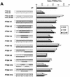
siRNA duplexes with a 3′ or 5′ overhang or without overhang (blunt) are equally potent in mediating gene silencing in HeLa cells. (A) Inhibition of PTEN mRNA expression in HeLa cells transfected with the indicated amounts of siRNA molecules. The sequences and different terminal structures of the siRNAs molecules are shown on the left. Mutations in the mismatch molecules are indicated by arrowheads. Samples were analysed in parallel for the level of PTEN mRNA expression 24 h after transfection by real time RT–PCR (Taqman) analysis. PTEN mRNA levels are shown relative to the mRNA levels of p110α, which served as internal reference. Each bar represents triplicate transfections (± SD). (B) Inhibition of PTEN protein expression by use of siRNAs with different terminal structures. The cells were harvested 48 h after transfection of the indicated siRNAs (30 nM) (lanes 2–7) or GeneBlocs (30 nM) (lanes 8 and 9). Cell extracts were separated by SDS–PAGE and analysed by immunoblotting using anti-p110α, anti-PTEN or anti-phospho-Akt antibody. The amount of p110α was used as a loading control and control cell extracts from untransfected HeLa cells (UT) were loaded in lane 1. (C) Inhibition of p110β mRNA expression in HeLa cells transfected with the indicated amounts of siRNA molecules. p110β mRNA levels are shown relative to the mRNA levels of p110α, which served as internal reference. The ratio of p110β/p110α mRNA of untransfected HeLa cells is shown at the bottom (UT). Each bar represents triplicate transfections (± SD).
Figure 3
Duplex length requirement and tolerance for mutation in siRNAs in HeLa cells. (A) Inhibition of Akt1 mRNA expression in HeLa cells transfected with the indicated amounts of siRNA molecules. The sequences, lengths and different terminal structures (3′ deoxyribonucleotides in upper case letters) of the siRNA molecules are shown on the left. The nucleotide changes in the mismatch siRNA molecule are indicated by arrowheads. Samples were analysed in parallel for the level of Akt1 and p110α mRNA expression 24 h after transfection by real time RT–PCR (Taqman) analysis. The mRNA levels of p110α served as internal reference. Each bar represents triplicate transfections (± SD). (B) Inhibition of PTEN mRNA expression in HeLa cells transfected with the indicated amounts of siRNA molecules. The sequences of the corresponding siRNA molecules are shown on the left. (C) Inhibition of PTEN protein expression analysed by immunoblot. The cells were harvested 48 or 96 h after transfection of the indicated siRNAs (30 nM). Cell extracts were separated by SDS–PAGE and analysed by immunoblotting as described previously. The positions of PTEN and p110α, which was used as a loading control, are indicated on the left.
Figure 4
RNAi activity and stability in serum of modified siRNA molecules. (A) Activity of siRNA with modified termini. 19mer duplex siRNAs specific for PTEN mRNA were synthesised with 2 nt deoxythymidine (TT) 3′ overhangs or without overhangs. iB represents inverted deoxy abasic end modifications, NH2 represents end protection with amino-C6 linker at the terminal phosphates. Inhibition of PTEN mRNA expression in HeLa cells transfected with the indicated amounts of modified siRNA molecules was determined by Taqman analysis as described before. (B) Stability assay of siRNAs with different chemical modifications. The structure of the modified siRNA molecules are schematically shown on the left. The siRNA molecule at the bottom was synthesized with 2′-_O_-methyl ribonucleotides (A, G, U and C) at all positions, indicated by IIIIII. Polyacrylamide gel (10%) electrophoresis of the indicated siRNA molecules after incubation in serum as described in Materials and Methods.
Figure 5
(Opposite) siRNA molecules with distinct 2′-_O_-methyl ribonucleotide modifications show increased stability in serum and mediate protein knock-down in HeLa cells. (A) RNAi activity of siRNA molecules with various 2′-_O_-methyl ribonucleotide modifications. Inhibition of PTEN mRNA expression in HeLa cells transfected with the indicated amounts of modified siRNA molecules. 2′-_O_-methyl ribonucleotide modifications are underlined and indicated by bold letters in the sequence on the left. Samples were analysed in parallel for the level of PTEN mRNA as described before. (B) Polyacrylamide gel electrophoresis of modified and unmodified siRNA molecules after incubation in serum. The PTEN siRNA sequence and the position of the 2′-_O_-methyl ribonucleotide modifications (underlined and bold letters) are shown on the left. (C) Inhibition of PTEN protein expression analysed by immunoblot. The cells were harvested 48 h after transfection of the indicated siRNAs (30 nM). Cell extracts were separated by SDS–PAGE and analysed by immunoblotting as described previously. The positions of PTEN and p110α, which was used as a loading control, are indicated at left. (D) siRNA molecules with distinct 2′-_O_-methyl ribonucleotide modifications mediate a prolonged protein knock-down. Inhibition of PTEN protein expression analysed by immunoblot. The cells were harvested 48 or 120 h after transfection of the indicated siRNAs (30 nM). Cell extracts were separated by SDS–PAGE and analysed by immunoblotting as described previously. The positions of PTEN and p110α, which was used as a loading control, are indicated on the left.
Figure 6
siRNA molecules with distinct 2′-_O_-methyl ribonucleotide modifications specific for Akt1 and p110β mRNA show increased stability in serum and mediate protein knock-down in HeLa cells. (A) The Akt1-specific siRNA sequence, the position of the 2′-_O_-methyl modifications (underlined and bold letters) and the integrity of the indicated siRNA molecules after incubation in serum are shown. (B) Inhibition of Akt1 protein expression as well as Akt phosphorylation analysed by immunoblot after transfection of the indicated siRNAs (30 nM). (C) Inhibition of the phosphorylation of the downstream kinase Akt1 by 2′-_O_-methyl-containing p110β-specific siRNAs. The positions of the phoshorylated Akt1 and p110α, which was used as a loading control, are indicated on the left.
Similar articles
- Positional effect of chemical modifications on short interference RNA activity in mammalian cells.
Prakash TP, Allerson CR, Dande P, Vickers TA, Sioufi N, Jarres R, Baker BF, Swayze EE, Griffey RH, Bhat B. Prakash TP, et al. J Med Chem. 2005 Jun 30;48(13):4247-53. doi: 10.1021/jm050044o. J Med Chem. 2005. PMID: 15974578 - Functional studies of the PI(3)-kinase signalling pathway employing synthetic and expressed siRNA.
Czauderna F, Fechtner M, Aygün H, Arnold W, Klippel A, Giese K, Kaufmann J. Czauderna F, et al. Nucleic Acids Res. 2003 Jan 15;31(2):670-82. doi: 10.1093/nar/gkg141. Nucleic Acids Res. 2003. PMID: 12527776 Free PMC article. - Effect of asymmetric terminal structures of short RNA duplexes on the RNA interference activity and strand selection.
Sano M, Sierant M, Miyagishi M, Nakanishi M, Takagi Y, Sutou S. Sano M, et al. Nucleic Acids Res. 2008 Oct;36(18):5812-21. doi: 10.1093/nar/gkn584. Epub 2008 Sep 9. Nucleic Acids Res. 2008. PMID: 18782830 Free PMC article. - Chemical modification of siRNA.
Deleavey GF, Watts JK, Damha MJ. Deleavey GF, et al. Curr Protoc Nucleic Acid Chem. 2009 Dec;Chapter 16:Unit 16.3. doi: 10.1002/0471142700.nc1603s39. Curr Protoc Nucleic Acid Chem. 2009. PMID: 20013783 Review. - [Nanostructured RNA for RNA interference].
Abe H. Abe H. Yakugaku Zasshi. 2013;133(3):373-8. doi: 10.1248/yakushi.12-00239-4. Yakugaku Zasshi. 2013. PMID: 23449417 Review. Japanese.
Cited by
- Mechanism of three-component collision to produce ultrastable pRNA three-way junction of Phi29 DNA-packaging motor by kinetic assessment.
Binzel DW, Khisamutdinov E, Vieweger M, Ortega J, Li J, Guo P. Binzel DW, et al. RNA. 2016 Nov;22(11):1710-1718. doi: 10.1261/rna.057646.116. Epub 2016 Sep 26. RNA. 2016. PMID: 27672132 Free PMC article. - In vivo screening of modified siRNAs for non-specific antiviral effect in a small fish model: number and localization in the strands are important.
Schyth BD, Bramsen JB, Pakula MM, Larashati S, Kjems J, Wengel J, Lorenzen N. Schyth BD, et al. Nucleic Acids Res. 2012 May;40(10):4653-65. doi: 10.1093/nar/gks033. Epub 2012 Jan 28. Nucleic Acids Res. 2012. PMID: 22287630 Free PMC article. - Effective inhibition of HIV-1 production by short hairpin RNAs and small interfering RNAs targeting a highly conserved site in HIV-1 Gag RNA is optimized by evaluating alternative length formats.
Scarborough RJ, Adams KL, Daher A, Gatignol A. Scarborough RJ, et al. Antimicrob Agents Chemother. 2015 Sep;59(9):5297-305. doi: 10.1128/AAC.00949-15. Epub 2015 Jun 15. Antimicrob Agents Chemother. 2015. PMID: 26077260 Free PMC article. - Leading RNA Interference Therapeutics Part 1: Silencing Hereditary Transthyretin Amyloidosis, with a Focus on Patisiran.
Titze-de-Almeida SS, Brandão PRP, Faber I, Titze-de-Almeida R. Titze-de-Almeida SS, et al. Mol Diagn Ther. 2020 Feb;24(1):49-59. doi: 10.1007/s40291-019-00434-w. Mol Diagn Ther. 2020. PMID: 31701435 Review. - Targeted delivery of siRNA.
Oliveira S, Storm G, Schiffelers RM. Oliveira S, et al. J Biomed Biotechnol. 2006;2006(4):63675. doi: 10.1155/JBB/2006/63675. J Biomed Biotechnol. 2006. PMID: 17057365 Free PMC article.
References
- Fire A., Xu,S., Montgomery,M.K., Kostas,S.A., Driver,S.E. and Mello,C.C. (1998) Potent and specific genetic interference by double-stranded RNA in Caenorhabditis elegans. Nature, 391, 806–811. - PubMed
- Hannon G.J. (2002) RNA interference. Nature, 418, 244–251. - PubMed
- Volpe T.A., Kidner,C., Hall,I.M., Teng,G., Grewal,S.I. and Martienssen,R.A. (2002) Regulation of heterochromatic silencing and histone H3 lysine-9 methylation by RNAi. Science, 297, 1833–1837. - PubMed
- Hall I.M., Shankaranarayana,G.D., Noma,K., Ayoub,N., Cohen,A. and Grewal,S.I. (2002) Establishment and maintenance of a heterochromatin domain. Science, 297, 2232–2237. - PubMed
Publication types
MeSH terms
Substances
LinkOut - more resources
Full Text Sources
Other Literature Sources
Research Materials


