Myoblasts transplanted into rat infarcted myocardium are functionally isolated from their host - PubMed (original) (raw)
Myoblasts transplanted into rat infarcted myocardium are functionally isolated from their host
Bertrand Leobon et al. Proc Natl Acad Sci U S A. 2003.
Abstract
Survival and differentiation of myogenic cells grafted into infarcted myocardium have raised the hope that cell transplantation becomes a new therapy for cardiovascular diseases. The approach was further supported by transplantation of skeletal myoblasts, which was shown to improve cardiac performance in several animal species. Despite the success of myoblast transplantation and its recent trial in human, the mechanism responsible for the functional improvement remains unclear. Here, we used intracellular recordings coupled to video and fluorescence microscopy to establish whether myoblasts, genetically labeled with enhanced GFP and transplanted into rat infarcted myocardium, retain excitable and contractile properties, and participate actively to cardiac function. Our results indicate that grafted myoblasts differentiate into peculiar hyperexcitable myotubes with a contractile activity fully independent of neighboring cardiomyocytes. We conclude that mechanisms other than electromechanical coupling between grafted and host cells are involved in the improvement of cardiac function.
Figures
Fig. 1.
Differences in dye coupling between transplanted myoblasts and host cells. (a) eGFP-expressing myotubes form bundles when transplanted in infarcted myocardium. A 2D projection of a stack of 20 images (each separated by 2 μm) obtained with two-photon microscopy. (b) Immunostaining of the heavy chain of the fast (Left) and the slow (Right) myosin isoforms. (c) A myotube expressing eGFP recorded intracellularly with an Alexa Fluor 568-containing micropipette (Left, FITC filters to reveal eGFP; Right, rhodamine filters to reveal Alexa Fluor). Thirty minutes after the impalement (Right), no dye coupling was observed between the myotube and neighboring myocytes. (d) A myocyte recorded intracellularly with an Alexa Fluor-containing micropipette (rhodamine filters). Labeling of the myocyte 30 s (Left) and 8 min (Right) after penetration. Dye coupling and labeling of neighboring myocytes occurred within 1–2 min. Scale bars = 20 μm.
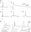
Fig. 2.
Emerging intrinsic membrane properties of eGFP-expressing myotubes. (a) Action potential characteristics of 5 types of muscle cell. (a1) Tibialis skeletal muscle cell; (a2) ventricular myocyte; (a3) eGFP-expressing myotube grafted for 28 days in the tibialis muscle; (a4) eGFP-expressing myotube grafted for 28 days in infarcted myocardium; (a5) eGFP-expressing myotube maintained in culture for 28 days. Whether grafted or in culture, eGFP-expressing myotubes fired brief action potentials (a3, a4, a5) in comparison with myocytes (a2). Note that the action potential of the myotubes grafted in infarcted myocardium (a4) is followed by a fast afterhyperpolarization. The action potentials were evoked either with extracellular stimulations (arrow heads point to the stimulation artifact) or with intracellular depolarizing current injections. (b) Firing properties of eGFP-expressing myotubes recorded in infarcted myocardium (Left and Center) or in culture (Right). (Left) Subthreshold depolarizing current pulses evoked a slow voltage-dependent depolarizing hump (arrow, middle trace) on top of which an action potential was triggered on higher stimulation (top trace). (Center) In another cell, a burst of action potentials was emitted on the slow depolarizing hump and followed by a slow afterhyperpolarization. (Right) Myotubes in culture do not fire bursts of action potentials.
Fig. 3.
Absence of electromechanical coupling between eGFP-expressing myotubes and myocytes. (Inset) Schematic representation of the experimental protocol used to detect contractions: movements of the recorded cell (red box) [i.e., an eGFP-expressing myotube (a and b) or a myocyte (c)] and of the neighboring myocardium (black box) were detected as local fluctuations in transilluminated light intensity. (a) Each action potential evoked in the myotube was accompanied by a local contraction (continuous red trace) that did not spread to the myocardium (continuous black trace). (b) The same myotube was electrically silent during spontaneous contractions of the entire explant. Light fluctuations detected in the red trace reflect only passive movements. (c) In opposition to the myotube, an intracellularly recorded myocyte fired with each contraction of the explant. The arrowheads point to the extracellular stimulations used to entrain the explant spontaneous contractions. The same calibration bars apply for a_–_c.
Similar articles
- Improvement of cardiac function in the failing rat heart after transfer of skeletal myoblasts engineered to overexpress placental growth factor.
Gmeiner M, Zimpfer D, Holfeld J, Seebacher G, Abraham D, Grimm M, Aharinejad S. Gmeiner M, et al. J Thorac Cardiovasc Surg. 2011 May;141(5):1238-45. doi: 10.1016/j.jtcvs.2010.10.054. Epub 2011 Feb 16. J Thorac Cardiovasc Surg. 2011. PMID: 21329947 - Endoventricular transplantation of allogenic skeletal myoblasts in a porcine model of myocardial infarction.
Dib N, Diethrich EB, Campbell A, Goodwin N, Robinson B, Gilbert J, Hobohm DW, Taylor DA. Dib N, et al. J Endovasc Ther. 2002 Jun;9(3):313-9. doi: 10.1177/152660280200900310. J Endovasc Ther. 2002. PMID: 12096946 - Skeletal myoblasts for cardiac repair: Act II?
Menasché P. Menasché P. J Am Coll Cardiol. 2008 Dec 2;52(23):1881-1883. doi: 10.1016/j.jacc.2008.07.066. J Am Coll Cardiol. 2008. PMID: 19038686 No abstract available. - Therapeutic potential of stem/progenitor cells in human skeletal muscle for cardiovascular regeneration.
Nomura T, Ashihara E, Tateishi K, Ueyama T, Takahas-Hi T, Yamagishi M, Kubo T, Yaku H, Matsubara H, Oh H. Nomura T, et al. Curr Stem Cell Res Ther. 2007 Dec;2(4):293-300. doi: 10.2174/157488807782793808. Curr Stem Cell Res Ther. 2007. PMID: 18220913 Review. - Skeletal myoblasts as a therapeutic agent.
Menasché P. Menasché P. Prog Cardiovasc Dis. 2007 Jul-Aug;50(1):7-17. doi: 10.1016/j.pcad.2007.02.002. Prog Cardiovasc Dis. 2007. PMID: 17631434 Review.
Cited by
- Stem cells for cardiac repair: an introduction.
du Pré BC, Doevendans PA, van Laake LW. du Pré BC, et al. J Geriatr Cardiol. 2013 Jun;10(2):186-97. doi: 10.3969/j.issn.1671-5411.2013.02.003. J Geriatr Cardiol. 2013. PMID: 23888179 Free PMC article. - Overexpression of connexin 43 using a retroviral vector improves electrical coupling of skeletal myoblasts with cardiac myocytes in vitro.
Tolmachov O, Ma YL, Themis M, Patel P, Spohr H, Macleod KT, Ullrich ND, Kienast Y, Coutelle C, Peters NS. Tolmachov O, et al. BMC Cardiovasc Disord. 2006 Jun 6;6:25. doi: 10.1186/1471-2261-6-25. BMC Cardiovasc Disord. 2006. PMID: 16756651 Free PMC article. - Stem cell therapies in cardiovascular disease A "realistic" appraisal.
Partovian C, Simons M. Partovian C, et al. Drug Discov Today Ther Strateg. 2008;5(1):73-78. doi: 10.1016/j.ddstr.2008.05.001. Drug Discov Today Ther Strateg. 2008. PMID: 19343101 Free PMC article. - Stem cells from in- or outside of the heart: isolation, characterization, and potential for myocardial tissue regeneration.
Noort WA, Sluijter JP, Goumans MJ, Chamuleau SA, Doevendans PA. Noort WA, et al. Pediatr Cardiol. 2009 Jul;30(5):699-709. doi: 10.1007/s00246-008-9370-5. Epub 2009 Jan 30. Pediatr Cardiol. 2009. PMID: 19184178 - The real estate of myoblast cardiac transplantation: negative remodeling is associated with location.
McCue JD, Swingen C, Feldberg T, Caron G, Kolb A, Denucci C, Prabhu S, Motilall R, Breviu B, Taylor DA. McCue JD, et al. J Heart Lung Transplant. 2008 Jan;27(1):116-23. doi: 10.1016/j.healun.2007.10.011. J Heart Lung Transplant. 2008. PMID: 18187097 Free PMC article.
References
- Menasche, P., Hagege, A. A., Scorsin, M., Pouzet, B., Desnos, M., Duboc, D., Schwartz, K., Vilquin, J. T. & Marolleau, J. P. (2001) Lancet 357, 279-280. - PubMed
- Taylor, D. A., Atkins, B. Z., Hungspreugs, P., Jones, T. R., Reedy, M. C., Hutcheson, K. A., Glower, D. D. & Kraus, W. E. (1998) Nat. Med. 4, 929-933. - PubMed
Publication types
MeSH terms
Substances
LinkOut - more resources
Full Text Sources
Other Literature Sources
Medical


