Activation of mitogen-activated protein kinase/extracellular signal-regulated kinase in hippocampal circuitry is required for consolidation and reconsolidation of recognition memory - PubMed (original) (raw)
Activation of mitogen-activated protein kinase/extracellular signal-regulated kinase in hippocampal circuitry is required for consolidation and reconsolidation of recognition memory
Aine Kelly et al. J Neurosci. 2003.
Abstract
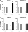
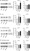
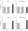
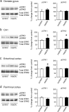
Consolidation and reconsolidation of long-term memory have been shown to be dependent on the synthesis of new proteins, but the specific molecular mechanisms underlying these events remain to be elucidated. The mitogen-activated protein kinase (MAPK) pathway can trigger genomic responses in neurons, leading to changes in protein synthesis, and several studies have identified its pivotal role in synaptic plasticity and long-term memory formation. In this study, we analyze the involvement of this pathway in the consolidation and reconsolidation of long-term recognition memory, using an object recognition task. We show that inhibition of the MAPK pathway by intracerebroventricular injection of the MEK [MAPK/extracellular signal-regulated kinase (ERK)] inhibitor UO126 blocks consolidation of object recognition memory but does not affect short-term memory. Brain regions of the entorhinal cortex-hippocampal circuitry were analyzed for ERK activation, and it was shown that consolidation of recognition memory was associated with increased phosphorylation of ERK in the dentate gyrus and entorhinal cortex, although total expression of ERK was unchanged. We also report that inhibition of the MAPK pathway blocks reconsolidation of recognition memory, and this was shown to be dependent on reactivation of the memory trace by brief reexposure to the objects. In addition, reconsolidation of memory was associated with an increase in the phosphorylation of ERK in entorhinal cortex and CA1. In summary, our data show that the MAPK kinase pathway is required for both consolidation and reconsolidation of long-term recognition memory, and that this is associated with hyperphosphorylation of ERK in different subregions of the entorhinal cortex-hippocampal circuitry.
Figures
Figure 1.
The effect of inhibition of MEK on consolidation of memory in object recognition. Arrows indicate injection of either a vehicle solution or the MEK inhibitor. Exploration of the objects in the sample phase are represented as light/dark gray bars. After the delay, exploration of the familiar object is represented by the white bar, and exploration of the novel object is represented by the black bar. a, Short-term retention. During the sample phase on day 1, rats spent an equivalent amount of time exploring the two objects. Exposure to a familiar and a novel object 10 min later resulted in significantly greater exploration of the novel object in both control and UO126-treated groups. b, Long-term retention. Both groups showed equal exploration of the two objects during the sample phase, but exposure to a familiar and a novel object 24 hr later on day 2 resulted in a significantly greater exploration of the novel object in the control group but not in the UO126-treated group. Histograms represent mean time in exploration with SEMs. Dashed lines represent equal exploration of the objects, and asterisks represent a significant increase in exploration of the object.
Figure 2.
An increase in ERK phosphorylation is associated with memory consolidation. Sample Western blots for each brain region are presented to the left of the histograms. a, Histograms show significant hyperphosphorylation of ERK1 in rats exploring the objects compared with control and naive rats with no change in pERK2 in the dentate gyrus. b, In dorsal CA1, pERK1 was significantly increased in the control rats and those exploring the objects compared with naive rats, with no change in pERK2. c, In the entorhinal cortex, although it did not reach statistical significance, there was a substantial increase in phosphorylation of both ERK1 and ERK2. d, No change in the phosphorylation of ERK1 and ERK2 was observed in the perirhinal cortex. No change in total ERK1 and ERK2 was observed in any group in any of the brain structures, asillustrated in the sample Western blots. Dotted lines represent control levels of pERK, and asterisks represent significant increase in pERK.
Figure 3.
a, The effect of MEK inhibition on the reconsolidation of memory. Arrows indicate injection of DMSO or UO126 on day 2. Graphical representation is the same as in Figure 1. Rats spent an equal amount of time exploring the two objects on both day 1 (D 1; sample phase) and day 2 (D 2; reactivation). Exposure to a familiar and a novel object on day 3 (D 3) resulted in a significantly greater exploration of the novel object in the control group but not in the UO126-treated group. b, Blockade of memory reconsolidation by MEK inhibition is dependent on reexposure to the objects. Both groups of rats spent an equal amount of time exploring the two objects on day 1. On day 2, rats received an injection of DMSO or UO126, as indicated by an arrow, but remained in the home cage. Exposure to a familiar and a novel object on day 3 resulted in a significantly greater exploration of the novel object in the control group in both control and UO126-treated groups. Dashed lines, SEMs, and asterisks are as referred to in Figure 1.
Figure 4.
An increase in ERK phosphorylation is associated with memory reconsolidation. Sample Western blots for each brain region are presented to the left of the histograms. Densitometric analysis was conducted on rats that were reexposed to the objects on day 2 to reactivate the memory trace (reactiv.), or control rats. a, In the dorsal dentate gyrus, there was no significant increase in pERK1 or pERK2 in the reactivation group. b, In dorsal CA1, there was a significant increase in pERK1 and pERK2 in the reactivation group. c, In entorhinal cortex, phosphorylation of ERK1 was significantly increased after reactivation compared with controls. d, No change in phosphorylation of either isoform of ERK was observed in the perirhinal cortex. No change in total ERK1 and ERK2 was observed in any group in any of the brain structures, as illustrated in thesample Western blots. Dotted lines and asterisks are as referred to in Figure 2.
Similar articles
- Differential BDNF signaling in dentate gyrus and perirhinal cortex during consolidation of recognition memory in the rat.
Callaghan CK, Kelly ÁM. Callaghan CK, et al. Hippocampus. 2012 Nov;22(11):2127-35. doi: 10.1002/hipo.22033. Epub 2012 May 10. Hippocampus. 2012. PMID: 22573708 - PKC-epsilon activation is required for recognition memory in the rat.
Zisopoulou S, Asimaki O, Leondaritis G, Vasilaki A, Sakellaridis N, Pitsikas N, Mangoura D. Zisopoulou S, et al. Behav Brain Res. 2013 Sep 15;253:280-9. doi: 10.1016/j.bbr.2013.07.036. Epub 2013 Jul 30. Behav Brain Res. 2013. PMID: 23911427 - The MAP(K) of fear: from memory consolidation to memory extinction.
Cestari V, Rossi-Arnaud C, Saraulli D, Costanzi M. Cestari V, et al. Brain Res Bull. 2014 Jun;105:8-16. doi: 10.1016/j.brainresbull.2013.09.007. Epub 2013 Sep 27. Brain Res Bull. 2014. PMID: 24080449 Review. - MAPK, CREB and zif268 are all required for the consolidation of recognition memory.
Bozon B, Kelly A, Josselyn SA, Silva AJ, Davis S, Laroche S. Bozon B, et al. Philos Trans R Soc Lond B Biol Sci. 2003 Apr 29;358(1432):805-14. doi: 10.1098/rstb.2002.1224. Philos Trans R Soc Lond B Biol Sci. 2003. PMID: 12740127 Free PMC article. Review.
Cited by
- NAc Shell Arc/Arg3.1 Protein Mediates Reconsolidation of Morphine CPP by Increased GluR1 Cell Surface Expression: Activation of ERK-Coupled CREB is Required.
Lv XF, Sun LL, Cui CL, Han JS. Lv XF, et al. Int J Neuropsychopharmacol. 2015 Mar 6;18(9):pyv030. doi: 10.1093/ijnp/pyv030. Int J Neuropsychopharmacol. 2015. PMID: 25746394 Free PMC article. - Systemic LPS induces spinal inflammatory gene expression and impairs phrenic long-term facilitation following acute intermittent hypoxia.
Huxtable AG, Smith SM, Vinit S, Watters JJ, Mitchell GS. Huxtable AG, et al. J Appl Physiol (1985). 2013 Apr;114(7):879-87. doi: 10.1152/japplphysiol.01347.2012. Epub 2013 Jan 17. J Appl Physiol (1985). 2013. PMID: 23329821 Free PMC article. - Proteomic and transcriptomic analysis of visual long-term memory in Drosophila melanogaster.
Jiang H, Hou Q, Gong Z, Liu L. Jiang H, et al. Protein Cell. 2011 Mar;2(3):215-22. doi: 10.1007/s13238-011-1019-0. Epub 2011 Apr 1. Protein Cell. 2011. PMID: 21461680 Free PMC article. - The extracellular signal-regulated kinase 1/2 pathway in neurological diseases: A potential therapeutic target (Review).
Sun J, Nan G. Sun J, et al. Int J Mol Med. 2017 Jun;39(6):1338-1346. doi: 10.3892/ijmm.2017.2962. Epub 2017 Apr 21. Int J Mol Med. 2017. PMID: 28440493 Free PMC article. Review.
References
- Abel T, Lattal KM ( 2001) Molecular mechanisms of memory acquisition, consolidation and retrieval. Curr Opin Neurobiol 11: 180–187. - PubMed
- Atkins CM, Selcher JC, Petraitis JJ, Trzaskos JM, Sweatt JD ( 1998) The MAPK cascade is required for mammalian associative learning. Nat Neurosci 1: 602–609. - PubMed
- Bradford M ( 1976) A rapid and sensitive method for the quantitation of microgram quantities of protein utilising the principle of protein dye binding. Anal Biochem 72: 248–254. - PubMed
Publication types
MeSH terms
Substances
LinkOut - more resources
Full Text Sources
Medical
Miscellaneous


