Increased cell proliferation and neurogenesis in the adult human Huntington's disease brain - PubMed (original) (raw)
Increased cell proliferation and neurogenesis in the adult human Huntington's disease brain
Maurice A Curtis et al. Proc Natl Acad Sci U S A. 2003.
Abstract
Neurogenesis has recently been observed in the adult human brain, suggesting the possibility of endogenous neural repair. However, the augmentation of neurogenesis in the adult human brain in response to neuronal cell loss has not been demonstrated. This study was undertaken to investigate whether neurogenesis occurs in the subependymal layer (SEL) adjacent to the caudate nucleus in the human brain in response to neurodegeneration of the caudate nucleus in Huntington's disease (HD). Postmortem control and HD human brain tissue were examined by using the cell cycle marker proliferating cell nuclear antigen (PCNA), the neuronal marker beta III-tubulin, and the glial cell marker glial fibrillary acidic protein (GFAP). We observed a significant increase in cell proliferation in the SEL in HD compared with control brains. Within the HD group, the degree of cell proliferation increased with pathological severity and increasing CAG repeats in the HD gene. Most importantly, PCNA+ cells were shown to coexpress beta III-tubulin or GFAP, demonstrating the generation of neurons and glial cells in the SEL of the diseased human brain. Our results provide evidence of increased progenitor cell proliferation and neurogenesis in the diseased adult human brain and further indicate the regenerative potential of the human brain.
Figures
Fig. 1.
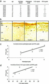
PCNA immunoreactivity is increased in the subependymal layer in HD cases. (a) Table summarizing the age, postmortem delay, pathological grade, CAG trinucleotide repeat length in the IT15 gene, and PCNA grade for each HD case is shown. For each HD case, the level of PCNA immunoreactivity in the subependymal layer was assessed by using a qualitative five-point grading scale (PCNA grade). (b) Compared with the control brain, the thickness of the subependymal layer (SEL) and the number of PCNA+ cells in the SEL are increased as the pathological grade of HD increases (grade 1 to grade 3). In each case, the boundary of the SEL with the caudate nucleus (CN) is indicated by an arrow. (EP, ependymal layer; LV, lateral ventricle). (c) Graph showing a significant correlation between pathological grade and PCNA grade in the HD cases (Spearman rank correlation,r = 0.8972, P ≤ 0.003). (d) Graph showing a significant correlation between CAG trinucleotide repeat length in the IT15 gene and PCNA grade in the HD cases (Spearman rank correlation, r = 0.8168, P ≤ 0.02). (Scale bar, 80 μm.)
Fig. 2.
Western immunoblotting with the PCNA antibody using human control (three cases) and HD (four cases) SEL homogenates demonstrates the specificity of the PCNA antibody by a single strong band at 36 kDa.
Fig. 3.
PCNA+, βIII-tubulin+, and GFAP+ cells are located in different regions within the SEL as demonstrated with bright-field immunohistochemical serial sections through the EP, SEL, and CN of a grade 2 HD brain. (a) PCNA+ cells are evenly distributed within the SEL (arrows demonstrate examples of PCNA+ cell bodies), and the EP is also densely stained. (b) βIII-tubulin+ fibers and cell bodies are present in the lower part of the SEL (arrows indicate cell bodies with immunoreactive fibers). (c) GFAP+ cells are distributed homogenously throughout the SEL and in the CN (arrows indicate examples of GFAP cell bodies).
Fig. 4.
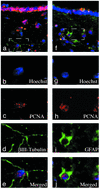
Newly generated cells in the SEL of the HD brain exhibit either a neuronal or glial phenotype. (a) Confocal microscopy demonstrates that the SEL of the HD brain contains newly generated cells that coexpress PCNA and βIII-tubulin and stain with a Hoechst stain. The new neurons are located in the lower part of the SEL. (Scale bar, 6 μm.) (b–d) Higher magnification of the neuron, which is outlined with a box in_a._ (Scale bar, 3 μm.) (b) Hoechst (blue) stains the nucleus of cells in the SEL. (c) PCNA (red) labels the nucleus of a new cell and has a granular appearance (19). (d) βIII-tubulin (green) labels the cytoplasm of neurons early in their development. (e) The merged image demonstrates coexpression of PCNA and βIII-tubulin in the same cell that displays a Hoechst+ stain, indicating that neurogenesis occurs in the SEL of the HD brain. (f) Confocal microscopy demonstrates that the SEL of the HD brain also contains newly generated cells that coexpress PCNA and GFAP and stain with a Hoechst stain. These new glial cells are located in the upper part of the SEL in the HD brain. (Scale bar, 18 μm.) (g–i) Higher magnification of the glial cell, which is outlined with a box in f. (Scale bar, 9 μm.) (g) Hoechst (blue) stains the nucleus of cells in the SEL. (h) PCNA (red) labels the nucleus of the cell with punctate staining. (i) The astrocytic marker GFAP (green) labels the cytoplasm of the cell. (j) The merged image demonstrates coexpression of PCNA and GFAP in the same cell that displays a Hoechst stain, indicating the occurrence of gliogenesis. Images a–e and_f–j_ were each obtained by using different excitation wavelengths, and the signal was detected at different emission wavelengths.
Similar articles
- The distribution of progenitor cells in the subependymal layer of the lateral ventricle in the normal and Huntington's disease human brain.
Curtis MA, Penney EB, Pearson J, Dragunow M, Connor B, Faull RL. Curtis MA, et al. Neuroscience. 2005;132(3):777-88. doi: 10.1016/j.neuroscience.2004.12.051. Neuroscience. 2005. PMID: 15837138 - A novel population of progenitor cells expressing cannabinoid receptors in the subependymal layer of the adult normal and Huntington's disease human brain.
Curtis MA, Faull RL, Glass M. Curtis MA, et al. J Chem Neuroanat. 2006 Apr;31(3):210-5. doi: 10.1016/j.jchemneu.2006.01.005. Epub 2006 Mar 14. J Chem Neuroanat. 2006. PMID: 16533591 - Activating transcription factor 2 expression in the adult human brain: association with both neurodegeneration and neurogenesis.
Pearson AG, Curtis MA, Waldvogel HJ, Faull RL, Dragunow M. Pearson AG, et al. Neuroscience. 2005;133(2):437-51. doi: 10.1016/j.neuroscience.2005.02.029. Neuroscience. 2005. PMID: 15878807 - Neurogenesis in Huntington's disease: can studying adult neurogenesis lead to the development of new therapeutic strategies?
Gil-Mohapel J, Simpson JM, Ghilan M, Christie BR. Gil-Mohapel J, et al. Brain Res. 2011 Aug 11;1406:84-105. doi: 10.1016/j.brainres.2011.06.040. Epub 2011 Jun 23. Brain Res. 2011. PMID: 21742312 Review. - Intervention of Proliferation and Differentiation of Endogenous Neural Stem Cells in the Neurodegenerative Process of Huntington's Disease Phenotype.
Mazurová Y, Gunčová I, Látr I, Rudolf E. Mazurová Y, et al. CNS Neurol Disord Drug Targets. 2011 Jun;10(4):486-99. doi: 10.2174/187152711795563967. CNS Neurol Disord Drug Targets. 2011. PMID: 21495959 Review.
Cited by
- An integrative systems-biology approach defines mechanisms of Alzheimer's disease neurodegeneration.
Leventhal MJ, Zanella CA, Kang B, Peng J, Gritsch D, Liao Z, Bukhari H, Wang T, Pao PC, Danquah S, Benetatos J, Nehme R, Farhi S, Tsai LH, Dong X, Scherzer CR, Feany MB, Fraenkel E. Leventhal MJ, et al. bioRxiv [Preprint]. 2024 Oct 9:2024.03.17.585262. doi: 10.1101/2024.03.17.585262. bioRxiv. 2024. PMID: 38559190 Free PMC article. Preprint. - Thyroid hormone action in adult neurogliogenic niches: the known and unknown.
Valcárcel-Hernández V, Mayerl S, Guadaño-Ferraz A, Remaud S. Valcárcel-Hernández V, et al. Front Endocrinol (Lausanne). 2024 Mar 7;15:1347802. doi: 10.3389/fendo.2024.1347802. eCollection 2024. Front Endocrinol (Lausanne). 2024. PMID: 38516412 Free PMC article. Review. - New implications for prion diseases therapy and prophylaxis.
Liu F, Lü W, Liu L. Liu F, et al. Front Mol Neurosci. 2024 Mar 4;17:1324702. doi: 10.3389/fnmol.2024.1324702. eCollection 2024. Front Mol Neurosci. 2024. PMID: 38500676 Free PMC article. Review. - The impact of adult neurogenesis on affective functions: of mice and men.
Alonso M, Petit AC, Lledo PM. Alonso M, et al. Mol Psychiatry. 2024 Aug;29(8):2527-2542. doi: 10.1038/s41380-024-02504-w. Epub 2024 Mar 18. Mol Psychiatry. 2024. PMID: 38499657 Free PMC article. Review. - Single-Nucleus RNA-Seq Characterizes the Cell Types Along the Neuronal Lineage in the Adult Human Subependymal Zone and Reveals Reduced Oligodendrocyte Progenitor Abundance with Age.
Puvogel S, Alsema A, North HF, Webster MJ, Weickert CS, Eggen BJL. Puvogel S, et al. eNeuro. 2024 Mar 4;11(3):ENEURO.0246-23.2024. doi: 10.1523/ENEURO.0246-23.2024. Print 2024 Mar. eNeuro. 2024. PMID: 38351133 Free PMC article.
References
- Svendsen, C. N., Caldwell, M. A., Shen, J., ter Borg, M. G., Rosser, A. E., Tyers, P., Karmiol, S. & Dunnett, S. B. (1997) Exp. Neurol. 148, 135-146. - PubMed
- Freed, C. R., Greene, P. E., Breeze, R. E., Tsai, W. Y., DuMouchel, W., Kao, R., Dillon, S., Winfield, H., Culver, S., Trojanowski, J. Q., et al. (2001) N. Engl. J. Med. 344, 710-719. - PubMed
- Morshead, C. M., Reynolds, B. A., Craig, C. G., McBurney, M. W., Staines, W. A., Morassutti, D., Weiss, S. & van der Kooy, D. (1994) Neuron 13, 1071-1082. - PubMed
- Arvidsson, A., Collin, T., Kirik, D., Kokaia, Z. & Lindvall, O. (2002) Nat. Med. 8, 963-970. - PubMed
Publication types
MeSH terms
Substances
LinkOut - more resources
Full Text Sources
Other Literature Sources
Medical
Miscellaneous


