Requirement of the Yersinia pseudotuberculosis effectors YopH and YopE in colonization and persistence in intestinal and lymph tissues - PubMed (original) (raw)
Requirement of the Yersinia pseudotuberculosis effectors YopH and YopE in colonization and persistence in intestinal and lymph tissues
Lauren K Logsdon et al. Infect Immun. 2003 Aug.
Abstract
The gram-negative enteric pathogen Yersinia pseudotuberculosis employs a type III secretion system and effector Yop proteins that are required for virulence. Mutations in the type III secretion-translocation apparatus have been shown to cause defects in colonization of the murine cecum, suggesting roles for one or more effector Yops in the intestinal tract. To investigate this possibility, isogenic yop mutant strains were tested for their ability to colonize and persist in intestinal and associated lymph tissues of the mouse following orogastric inoculation. In single-strain infections, a yopHEMOJ mutant strain was unable to colonize, replicate, or persist in intestinal and lymph tissues. A yopH mutant strain specifically fails to colonize the mesenteric lymph nodes, but yopE and yopO mutant strains showed only minor defects in persistence in intestinal and lymph tissues. While no single Yop was found to be essential for colonization or persistence in intestinal tissues in single-strain infections, the absence of both YopH and YopE together almost eliminated colonization of all tissues, indicating either that these two Yops have some redundant functions or that Y. pseudotuberculosis employs multiple strategies for colonization. In competition infections with wild-type Y. pseudotuberculosis, the presence of wild-type bacteria severely hindered the ability of the yopH, yopE, and yopO mutants to persist in many tissues, suggesting that the wild-type bacteria either fills colonization niches or elicits host responses that the yop mutants are unable to withstand.
Figures
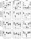
FIG. 1.
Colonization of intestinal and lymph tissues at day 1 and day 2 postinfection with 2 × 10⁹ CFU of wild-type Y. pseudotuberculosis (filled diamonds), yopHEMOJ (open circles), or yopBHEMOJ (open triangles). Data from four to six mice from two experiments were pooled. Each symbol represents the log10 CFU/gram of tissue from one mouse colonized with the appropriate strain. All points were above the limit of detection of 10 CFU/gram of tissue. At day 1, colonization by yopHEMOJ and yopBHEMOJ was statistically different (**; P < 0.01) from that by the wild-type strain in the ileum, colon, and PP. At day 2, colonization by yopHEMOJ and yopBHEMOJ are statistically different from the wild-type strain (P < 0.01) in all tissues.
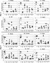
FIG. 2.
Colonization of ileum, PP, and MLN at day 2 and day 5 postinfection with either 2 × 10⁹ CFU or 2 × 10⁸ CFU of wild-type (wt), yopB (ΔB), yopH (ΔH), yopE (ΔE), or yopO (ΔO) Y. pseudotuberculosis. Data from four to eight mice from two to four experiments were pooled. Each symbol represents the log10 CFU/gram of tissue for the tissue sample from one mouse. Shaded bars represent the average log10 CFU/gram of tissue. Open symbols indicate that less than 10 CFU were recovered/tissue. Asterisks indicate statistical significance of difference between colonization levels of yop mutants and wild-type bacteria as follows: *, P = 0.1 to 0.01; **, P < 0.01.
FIG. 3.
Colonization of the spleen at 2 and 5 days postinfection with either 2 × 10⁹ CFU or 2 × 10⁸ CFU of wild-type (wt), yopB (ΔB), yopH (ΔH), yopE (ΔE), yopO (ΔO) or ΔHEMOJ Y. pseudotuberculosis. Data from four to eight mice from two to four experiments were pooled. Each symbol represents the log10 CFU/gram of tissue for the spleen from one mouse. Open symbols indicate that less than 10 CFU/tissue were recovered.
FIG. 4.
Colonization of intestinal and lymph tissues at day 2 and day 5 postinfection with 2 × 10⁸ CFU of wild-type, yopHE (ΔHE), yopHO (ΔHO), yopEO (ΔEO), yopHEO (ΔHEO), or yopHEMOJ (ΔHEMOJ) Y. pseudotuberculosis. Data from four to six mice from two to three experiments were pooled. Each symbol represents the log10 CFU/gram of tissue for the tissue sample from one mouse. Shaded bars represent the average log10 CFU/gram of tissue. Open symbols indicate that less than 10 CFU were recovered/tissue. Asterisks indicate statistical significance of difference between colonization levels of yop mutants and wild-type bacteria. *, P = 0.1 to 0.01; **, P < 0.01.
FIG. 5.
Competition of yopHE versus yopHEMOJ (closed circles) and yopHEO versus yopHEMOJ (open circles), 1 day postinfection with 2 × 10⁹ CFU of an equal mixture of yopHE and yopHEMOJ or yopHEO and yopHEMOJ. Data from three to six mice from two experiments were pooled. Data are graphed as C.I. {[yopHE(O)/yopHEMOJ output ratio]/[yopHE(O)/yopHEMOJ input ratio]} values for the tissue samples from one mouse. Shaded bars represent geometric means of the C.I. values. All points were above the limit of detection.
FIG. 6.
Competition between wild-type Y. pseudotuberculosis and yop mutants at 5 days postinfection with 2 × 10⁹ CFU of a mixture of equal amounts of the wild-type strain and either yopB, yopH, yopE, yopO, or yopHEMOJ. Data from six to eight mice from three to four experiments were pooled. Each point represents the C.I. [(mutant/wild-type output ratio)/(mutant/wild-type input ratio)] value for the tissue sample from one mouse. Shaded bars represent the geometric means of the C.I. values. Open symbols indicate that the yop mutant was below the limit of detection, which was usually 200 bacteria. Asterisks indicate the yop mutants that were significantly outcompeted by wild-type bacteria (*, P = 0.1 to 0.01; **, P < 0.01).
Similar articles
- Yersinia pseudotuberculosis efficiently escapes polymorphonuclear neutrophils during early infection.
Westermark L, Fahlgren A, Fällman M. Westermark L, et al. Infect Immun. 2014 Mar;82(3):1181-91. doi: 10.1128/IAI.01634-13. Epub 2013 Dec 30. Infect Immun. 2014. PMID: 24379291 Free PMC article. - The Yersinia Yop virulon: a bacterial system for subverting eukaryotic cells.
Cornelis GR, Wolf-Watz H. Cornelis GR, et al. Mol Microbiol. 1997 Mar;23(5):861-7. doi: 10.1046/j.1365-2958.1997.2731623.x. Mol Microbiol. 1997. PMID: 9076724 Review. - The Yersinia Yop virulon, a bacterial system to subvert cells of the primary host defense.
Cornelis GR. Cornelis GR. Folia Microbiol (Praha). 1998;43(3):253-61. doi: 10.1007/BF02818610. Folia Microbiol (Praha). 1998. PMID: 9717252 Review.
Cited by
- ROS-inhibitory activity of YopE is required for full virulence of Yersinia in mice.
Songsungthong W, Higgins MC, Rolán HG, Murphy JL, Mecsas J. Songsungthong W, et al. Cell Microbiol. 2010 Jul;12(7):988-1001. doi: 10.1111/j.1462-5822.2010.01448.x. Epub 2010 Feb 9. Cell Microbiol. 2010. PMID: 20148901 Free PMC article. - Early apoptosis of macrophages modulated by injection of Yersinia pestis YopK promotes progression of primary pneumonic plague.
Peters KN, Dhariwala MO, Hughes Hanks JM, Brown CR, Anderson DM. Peters KN, et al. PLoS Pathog. 2013;9(4):e1003324. doi: 10.1371/journal.ppat.1003324. Epub 2013 Apr 25. PLoS Pathog. 2013. PMID: 23633954 Free PMC article. - Hfq regulates biofilm gut blockage that facilitates flea-borne transmission of Yersinia pestis.
Rempe KA, Hinz AK, Vadyvaloo V. Rempe KA, et al. J Bacteriol. 2012 Apr;194(8):2036-40. doi: 10.1128/JB.06568-11. Epub 2012 Feb 10. J Bacteriol. 2012. PMID: 22328669 Free PMC article. - Contribution of the major secreted yops of Yersinia enterocolitica O:8 to pathogenicity in the mouse infection model.
Trülzsch K, Sporleder T, Igwe EI, Rüssmann H, Heesemann J. Trülzsch K, et al. Infect Immun. 2004 Sep;72(9):5227-34. doi: 10.1128/IAI.72.9.5227-5234.2004. Infect Immun. 2004. PMID: 15322017 Free PMC article.
References
- Andersson, K., N. Carballeira, K. E. Magnusson, C. Persson, O. Stendahl, H. Wolf-Watz, and M. Fallman. 1996. YopH of Yersinia pseudotuberculosis interrupts early phosphotyrosine signalling associated with phagocytosis. Mol. Microbiol. 20:1057-1069. - PubMed
- Autenrieth, I. B., and R. Firsching. 1996. Penetration of M cells and destruction of Peyer's patches by Yersinia enterocolitica: an ultrastructural and histological study. J. Med. Microbiol. 44:285-294. - PubMed
- Barz, C., T. N. Abahji, K. Trulzsch, and J. Heesemann. 2000. The Yersinia Ser/Thr protein kinase YpkA/YopO directly interacts with the small GTPases RhoA and Rac-1. FEBS Lett. 482:139-143. - PubMed
- Black, D. S., and J. B. Bliska. 2000. The RhoGAP activity of the Yersinia pseudotuberculosis cytotoxin YopE is required for antiphagocytic function and virulence. Mol. Microbiol. 37:515-527. - PubMed
Publication types
MeSH terms
Substances
Grants and funding
- P30 DK34928/DK/NIDDK NIH HHS/United States
- P30 DK034928/DK/NIDDK NIH HHS/United States
- T32-AI 07422/AI/NIAID NIH HHS/United States
- T32 AI007422/AI/NIAID NIH HHS/United States
- R21-AI49348/AI/NIAID NIH HHS/United States
LinkOut - more resources
Full Text Sources
Other Literature Sources


