Neuronal migration from the forebrain to the olfactory bulb requires a new attractant persistent in the olfactory bulb - PubMed (original) (raw)
Neuronal migration from the forebrain to the olfactory bulb requires a new attractant persistent in the olfactory bulb
Guofa Liu et al. J Neurosci. 2003.
Abstract
Interneurons in the olfactory bulb (OB) are generated not only in the developing embryo but also throughout the postnatal life of mammals from neuronal precursor cells migrating from the anterior subventricular zone (SVZa) of the mammalian forebrain. We discovered that the OB secretes a diffusible activity that attracts these neuronal precursor cells. The attractive activity is present in specific layers in the OB, including the glomerular layer but not the granule cell layer. The attractive activity and the neuronal responsiveness persist from embryonic through neonatal to adult stages. Removal of the rostral OB significantly reduces SVZa migration toward the OB, an effect that can be rescued by a transplant of the OB but not by that of the neocortex. The activity in the OB is not mimicked by the known attractants. These results provide an explanation for the continuous migration of SVZa neurons toward the OB, demonstrate an important role of the OB in neuronal migration, and reveal the existence of a new chemoattractant.
Figures
Figure 1.
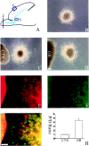
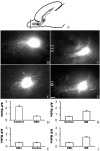
Presence of a chemoattractive activity in the OB. A, A diagram of the sagittal section of neonatal rat forebrain. X, SVZa; Y, region of neocortex from which cortical explants were isolated and used in C; Z, tip of the OB from which explants were isolated and used in D-G. B, An SVZa explant was cultured for 24 hr. Cells migrated symmetrically from the explant. C, Coculture of an SVZa explant with a neocortical explant (CTX). Migrating SVZa cells were symmetrically distributed around the circumference of the SVZa explant (n = 129 of 137). D, Effect of the OB on the migration of cells from SVZa explants. Asymmetric distribution of migrating cells with a higher number of cells in the quadrant proximal to the OB than that in distal quadrant (n = 539 of 627) is shown. Scale bar, 100 μm. E, The same explant as in D, showing staining with the TuJ1 antibody. F, The same explant as in D, showing staining with anti-GABA antibodies. G, Superimposition of E and F. H, Effects of cortical and OB explants on the distribution of SVZa cell migration. Proximal/distal ratios were calculated from the numbers of cells in the proximal quadrants divided by those in the distal quadrants. Cell numbers were counted from 33 cocultures with the OB explants and 16 cortical (CTX) explants. CTX, Cortex.
Figure 2.
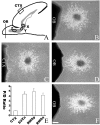
Attractive effects of the OB on cells migrating along the RMS. A, A diagram of the sagittal section of neonatal rat forebrain showing different parts of the RMS from which explants were isolated and cocultured. X, SVZa; Y, posterior part of RMS; Z, anterior part of the RMS. B, Asymmetric distribution of cells migrating from an SVZa explant cocultured with the OB. C, Symmetric distribution of cells migrating from an SVZa explant cocultured with a cortical explant. D, Asymmetric distribution of cells migrating from an explant from the posterior part of the RMS cocultured with the OB (n = 236 of 291). E, Effects of cortical and OB explants on the distribution of cells migrating out of explants of the SVZa and RMS. The cell numbers were counted from 33 SVZa explants, 32 posterior RMS explants, and 36 anterior RMS explants. CTX, Cortex; RMSp, posterior RMS; RMSa, anterior RMS; P/D, proximal/distal. F, Asymmetric distribution of cells migrating from an anterior RMS explant cocultured with the OB (n = 312 of 372). Scale bar, 100 μm.
Figure 3.
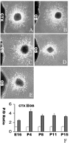
Persistent chemoattractive activity in the OB from embryonic to adult stages. A, Effect of an E16 rat cortical explant on cells migrating from a P4 SVZa explant. B, Effect of an E16 OB explant on cells migrating from a P4 SVZa explant. C, Effect of an E20 OB explant on cells migrating from a P4 SVZa explant. D, Effect of a P4 OB explant on cells migrating from a P4 SVZa explant. E, Effect of a P8 OB explant on cells migrating from a P4 SVZa explant. F, Effect of a P11 OB explant on cells migrating from a P4 SVZa explant. G, Effect of an adult OB explant on cells migrating from a P4 SVZa explant. Scale bar, 100 μm. H, The proximal/distal (P/D) ratios of cell numbers were counted from 21 cortical and 31 E16 OB explants of E16 rats, 25 cortical and 25 OB explants of E20 rats, 16 cortical and 33 OB explants of P4 rats, 26 cortical and 22 OB explants of P8 rats, 22 cortical and 19 OB explants of P11 rats, and 26 cortical and 25 OB explants of adult rats. The difference between cortical and OB explants was statistically very significant (p < 0.0001). CTX, Cortex.
Figure 4.
Persistent neuronal responsiveness to the OB chemoattractant(s). A, Effect of a P4 cortical explant on cells migrating from an E16 LGE explant. B, Effect of a P4 OB explant on cells migrating from an E16 LGE explant. C, Effect of a P4 OB explant on cells migrating from a P8 SVZa explant. D, Effect of a P4 OB explant on cells migrating from a P11 SVZa explant. E, Effect of a P4 OB on cells migrating from a P15 SVZa explant. Scale bar, 100 μm. F, The proximal/distal (P/D) ratios of cell numbers were counted from 25 P4 cortical and 21 P4 OB cocultures with E16 LGE explants, 16 cortical and 33 OB cocultures with P4 SVZa explants, 32 cortical and 30 OB cocultures with P8 SVZa explants, 24 cortical and 21 OB cocultures with P11 SVZa explants, and 25 cortical and 24 OB cocultures with P15 SVZa explants. The difference between cortical and OB cocultures was statistically very significant (p < 0.0001).
Figure 5.
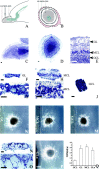
Dissection of OB layers with the chemoattractive activity. A, A diagram of the sagittal section of neonatal forebrain showing the anatomic layers of the OB. B, A diagram of the coronal section of the neonatal OB (outlined by dashed line). The layer between the GL and MCL is the external plexiform layer, whereas the layer between the MCL and GCL is the internal plexiform layer. C, A sagittal slice of the P4 OB stained with cresyl violet showing the different layers in the OB. D, A coronal slice of P4 OB stained with cresyl violet. E, A higher magnification view of C showing the GL, external plexiform layer, MCL, internal plexiform layer, and GCL. F, A GL explant dissected from a P4 OB was stained with cresyl violet. G, A higher magnification view of F showing that it contains GL. H, An explant dissected from a P4 OB was stained with cresyl violet, showing that it contains the external plexiform layer, MCL, and internal plexiform layer. I, A higher magnification view of H. J, A GCL explant from a P4 OB was stained with cresyl violet, showing that it contains only GCL but not the other layers in the OB. K, Effect of the GL explants on cells migrating from a P4 SVZa explant (n = 66 of 74). L, Effect of the MCL explants (including the MCL and parts of the external and internal plexiform layers) on cells migrating from a P4 SVZa explant (n = 58 of 61). M, Effect of GCL explants on cells migrating from a P4 SVZa explant (n = 69 of 77). N, A GL explant dissected from the adult OB was stained with cresyl violet, showing that it contains only the GL. O, A higher magnification view of N. P, Effect of an adult GL explant on cells migrating from a P4 SVZa explant (n = 87 of 93). GLa, Adult GL. Q, Effects of different layers from the OB on the distribution of cells migrating out of the SVZa explants. The cell numbers were counted from 24 P4 GCL explants, 26 P4 GL explants, 22 P4 MCL explants, and 25 adult GL (GLa) explants. Scale bars, 100 μm.
Figure 6.
An essential role of the OB in directing neuronal migration in the RMS. A, A diagram of the sagittal section of a neonatal forebrain showing the position of DiI insertion at the juncture of the anterior (A) and posterior (P) parts of the RMS. B, In a normal slice, more cells migrating anteriorly toward the OB. C, When the rostral end of the OB was removed, the distribution of migrating cells was changed with reduced anterior migration and increased posterior migration. D, Effect of OB removal in directing neuronal migration in the RMS. The cell numbers were counted from 20 intact sagittal slices and 32 sagittal slices without the rostral end of the OB. E, Cortical transplants could not functionally replace the rostral end of the OB in directing anterior migration. The cell numbers were counted from 32 slices with the rostral end of their OB removed and nine cortical transplants placed at the rostral end of the OB. F, When a cortical transplant was used to replace the rostral end of the OB, the distribution of migrating cells could not be rescued. G, When an OB transplant was placed at the rostral end of the OB after the original rostral end of the OB was removed, the distribution of migrating cells was changed with more cells migrating anteriorly toward the OB. H, I, The cell numbers were counted from 32 sagittal slices without the rostral end of the OB, nine cortical transplants, and seven OB transplants placed at the rostral end of the OB. CTX, Cortex; A/P, anterior/posterior. Scale bar, 100 μm.
Figure 7.
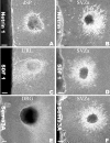
Inability of known chemoattractants to mimic the attractive activity in the OB. A, Chemoattraction of E12 commissural axons by netrin-1 expressed from a stable HEK cell line. dSP, Dorsal spinal cord. B, Chemorepulsion of SVZa cells by netrin-1 from the same HEK cell line as those used in A. C, Chemoattraction of E15 upper rhombic lip (URL) by SDF-1 expressed from a stable HEK cell line. D, Absence of guidance activity on SVZa neurons by SDF-1 from the same cell line as that in C. E, Chemorepulsion of E14 dorsal root ganglion (DRG) axon by Sema 3A transiently expressed from an HEK cell line. F, Absence of guidance activity on SVZa neurons by Sema 3A transiently expressed from an HEK cell line. Scale bar, 100 μm.
Similar articles
- IGF-I promotes neuronal migration and positioning in the olfactory bulb and the exit of neuroblasts from the subventricular zone.
Hurtado-Chong A, Yusta-Boyo MJ, Vergaño-Vera E, Bulfone A, de Pablo F, Vicario-Abejón C. Hurtado-Chong A, et al. Eur J Neurosci. 2009 Sep;30(5):742-55. doi: 10.1111/j.1460-9568.2009.06870.x. Epub 2009 Aug 27. Eur J Neurosci. 2009. PMID: 19712103 - The division of neuronal progenitor cells during migration in the neonatal mammalian forebrain.
Menezes JR, Smith CM, Nelson KC, Luskin MB. Menezes JR, et al. Mol Cell Neurosci. 1995 Dec;6(6):496-508. doi: 10.1006/mcne.1995.0002. Mol Cell Neurosci. 1995. PMID: 8742267 - Neuroblasts of the postnatal mammalian forebrain: their phenotype and fate.
Luskin MB. Luskin MB. J Neurobiol. 1998 Aug;36(2):221-33. J Neurobiol. 1998. PMID: 9712306 Review. - Dopamine systems in the forebrain.
Cave JW, Baker H. Cave JW, et al. Adv Exp Med Biol. 2009;651:15-35. doi: 10.1007/978-1-4419-0322-8_2. Adv Exp Med Biol. 2009. PMID: 19731547 Free PMC article. Review.
Cited by
- Subventricular zone cell migration: lessons from quantitative two-photon microscopy.
James R, Kim Y, Hockberger PE, Szele FG. James R, et al. Front Neurosci. 2011 Mar 21;5:30. doi: 10.3389/fnins.2011.00030. eCollection 2011. Front Neurosci. 2011. PMID: 21472025 Free PMC article. - Multiple roles for slits in the control of cell migration in the rostral migratory stream.
Nguyen-Ba-Charvet KT, Picard-Riera N, Tessier-Lavigne M, Baron-Van Evercooren A, Sotelo C, Chédotal A. Nguyen-Ba-Charvet KT, et al. J Neurosci. 2004 Feb 11;24(6):1497-506. doi: 10.1523/JNEUROSCI.4729-03.2004. J Neurosci. 2004. PMID: 14960623 Free PMC article. - N-syndecan deficiency impairs neural migration in brain.
Hienola A, Tumova S, Kulesskiy E, Rauvala H. Hienola A, et al. J Cell Biol. 2006 Aug 14;174(4):569-80. doi: 10.1083/jcb.200602043. J Cell Biol. 2006. PMID: 16908672 Free PMC article. - Control of neuronal migration through rostral migration stream in mice.
Sun W, Kim H, Moon Y. Sun W, et al. Anat Cell Biol. 2010 Dec;43(4):269-79. doi: 10.5115/acb.2010.43.4.269. Epub 2010 Dec 31. Anat Cell Biol. 2010. PMID: 21267400 Free PMC article. - An Overview of Adult Neurogenesis.
Ribeiro FF, Xapelli S. Ribeiro FF, et al. Adv Exp Med Biol. 2021;1331:77-94. doi: 10.1007/978-3-030-74046-7_7. Adv Exp Med Biol. 2021. PMID: 34453294 Review.
References
- Alcantara S, Ruiz M, Castro FD, Soriano E, Sotelo C ( 2000) Netrin 1 acts as an attractive or a repulsive cue for distant migrating neurons during the development of the cerebellar system. Development 127: 1359-1372. - PubMed
- Altman J ( 1969) Autoradiographic and histological studies of postnatal neurogenesis. IV. Cell proliferation and migration in the anterior forebrain with special reference to persisting neurogenesis in the olfactory bulb. J Comp Neurol 137: 433-457. - PubMed
- Altman J, Das GD ( 1966) Autoradiographic and histological studies of postnatal neurogenesis. I. A longitudinal investigation of the kinetics, migration and transformation of cells incorporating tritiated thymidine in neonate rats, with special reference to postnatal neurogenesis in some brain regions. J Comp Neurol 126: 337-389. - PubMed
- Alvarez-Buylla A ( 1997) Mechanism of migration of olfactory bulb interneurons. Cell Dev Biol 8: 207-213. - PubMed
- Axel R ( 1995) The molecular logic of smell. Sci Am 4: 154-159. - PubMed
Publication types
MeSH terms
Substances
LinkOut - more resources
Full Text Sources
Other Literature Sources
Medical
Miscellaneous
