Structural basis for the product specificity of histone lysine methyltransferases - PubMed (original) (raw)
Comparative Study
Structural basis for the product specificity of histone lysine methyltransferases
Xing Zhang et al. Mol Cell. 2003 Jul.
Abstract
DIM-5 is a SUV39-type histone H3 Lys9 methyltransferase that is essential for DNA methylation in N. crassa. We report the structure of a ternary complex including DIM-5, S-adenosyl-L-homocysteine, and a substrate H3 peptide. The histone tail inserts as a parallel strand between two DIM-5 strands, completing a hybrid sheet. Three post-SET cysteines coordinate a zinc atom together with Cys242 from the SET signature motif (NHXCXPN) near the active site. Consequently, a narrow channel is formed to accommodate the target Lys9 side chain. The sulfur atom of S-adenosyl-L-homocysteine, where the transferable methyl group is to be attached in S-adenosyl-L-methionine, lies at the opposite end of the channel, approximately 4 A away from the target Lys9 nitrogen. Structural comparison of the active sites of DIM-5, an H3 Lys9 trimethyltransferase, and SET7/9, an H3 Lys4 monomethyltransferase, allowed us to design substitutions in both enzymes that profoundly alter their product specificities without affecting their catalytic activities.
Figures
Figure 1. Domain Structure of SET HKMT Families
The DIM-5 protein (the smallest known member of the Suv39 family) contains four segments: a weakly conserved amino-terminal region (light blue), a pre-SET domain (yellow) containing nine invariant cysteines, the SET region (green) containing signature motifs NHXCXPN and ELXFDY (magenta), and the post-SET domain (gray) containing three invariant cysteines.
Figure 2. DIM-5 Kinetics and Ternary Structure
(A) Mass spectrometry analysis of different H3 peptides as DIM-5 substrates. The relative amount of each peptide species, expressed as a percentage of the sum of intensity of all related peaks, was plotted over the full time courses of the reactions. (B) GRASP (Nicholls et al., 1991) surface charge distribution (blue for positive, red for negative, white for neutral). The H3 peptide and AdoHcy are shown as stick models. (C) Ribbon (Carson, 1997) diagram colored as in Figure 1. The pre-SET residues (yellow) form a Zn3Cys9 triangular zinc cluster. The SET residues (green) and the N-terminal region are folded into six β sheets surrounding a knot-like structure (magenta). The post-SET residues (gray) bind the fourth zinc atom, adjacent to the substrate H3 peptide (red) and AdoHcy (blue). (D) The substrate H3 peptide (red), superimposed on an omit electron density contoured at 4.0 σ (orange), is inserted as a parallel β strand (red in Figure 2C) between two DIM-5 strands, β 10 (green) and β 18 (magenta). The side chain density for H3 Arg-8 is complete at lower contour levels (2.5 σ in Fobs-Fcal and 0.8 σ in 2Fobs-Fcal).
Figure 3. The Methylation Mechanism
(A) The post-SET zinc ion and the AdoHcy binding site. The zinc ion is presented as a red ball, coordinated by four cysteines, C244 (magenta) and C306XC308X4C313 (gray). AdoHcy is superimposed onto a difference electron density map contoured at 4.0 σ (orange). Dashed lines indicate the hydrogen bonds. One face of the AdoHcy adenine base lies against the aliphatic portion of R159, whose guanidino group forms a bifurcated salt bridge with the two carboxyl oxygen atoms of E278. The polar edge of the adenine base forms three hydrogen bonds to the DIM-5 backbone: the exocyclic amino group N6 with the carbonyl oxygen of H242, the ring N1 with the amide of L307, and the ring N7 with the amide of H242. The adenine ring carbon C8 makes van der Waals contacts to the Y283 hydroxyl and with the side chain carbonyl Oδ1 of N241; this explains the complete loss of AdoMet crosslinking in N241Q and Y283F mutants (Zhang et al., 2002). The two ribose hydroxyls interact with the main chain amide of V203 and the side chain carboxyl of D202. The amino group of the homocysteine moiety hydrogen bonds the side chain Oδ1 of N241, while its side chain amino group forms two hydrogen bonds with the backbone carbonyl of W161 and with the side chain of E278. The carboxylate group of the homocysteine moiety interacts with Y204 and the backbone amide of W161. (B) Close-up view of the H3 peptide binding site with Lys9 inserted into a channel. (C) The target Lys binding site (stereo). The arrow indicates the movement of the methyl group transferred from the AdoMet methylsulfonium group to the target amino group. (D) DIM-5 activity (LogCPM) as a function of pH. (E) AdoHcy bound in a large surface pocket, allowing for processive methylation. The green ellipse indicates the location where the AdoHcy homocysteine moiety binds in the peptide-free structure (Zhang et al., 2002).
Figure 4. Enzymatic Properties of Recombinant DIM-5 and SET7/9 Mutants
(A) Activities of DIM-5 and SET7/9 mutants using histone substrate (top). AdoMet crosslinking experiments of DIM-5 showing fluorograph (middle) and Coomassie stain (bottom). (B) Structure-based sequence alignment of DIM-5 and SET7/9. Secondary structures shown are based on Wilson et al. (2002) and Zhang et al. (2002). Vertical bars indicate residues that align spatially. Residues identical (black background) or similar (gray background) between the two enzymes, as well as the post-SET region of DIM-5, are highlighted. Numbered residues are described in the text. C-terminal hydrophobic residues of DIM-5 are underlined. (C) Structural comparison of active sties in the ternary DIM-5 (in color) and binary SET7/9-AdoHcy (in black) (PDB 1MT6; Jacobs et al., 2002). The bound peptide in DIM-5 is represented as a solid electron density (orange), with the target Lys surrounded by either two Tyr and one Phe (DIM-5) or three Tyr (SET7/9).
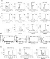
Figure 5. Mass Spectrometry Analysis of Methylation Kinetics
(A) Representative spectra at various time points for WT DIM-5, its F281Y variant, WT SET7/9, and its Y305F variant are shown. The peaks for unmodified (Um) substrate and mono-, di-, and trimethylated products are labeled. Unlabeled minor peaks correspond to the sodium adducts of the major peaks (+23 Da). (B) The relative amount of each peptide species over the full time courses of the reactions, expressed as a percentage of the sum of intensity of all related peaks. (C) Spectra for three DIM-5 mutants having severely impaired catalytic activity but with normal product specificity.
Comment in
- Cracking the histone code: one, two, three methyls, you're out!
Dutnall RN. Dutnall RN. Mol Cell. 2003 Jul;12(1):3-4. doi: 10.1016/s1097-2765(03)00282-x. Mol Cell. 2003. PMID: 12887887 Review.
Similar articles
- Trimethylated lysine 9 of histone H3 is a mark for DNA methylation in Neurospora crassa.
Tamaru H, Zhang X, McMillen D, Singh PB, Nakayama J, Grewal SI, Allis CD, Cheng X, Selker EU. Tamaru H, et al. Nat Genet. 2003 May;34(1):75-9. doi: 10.1038/ng1143. Nat Genet. 2003. PMID: 12679815 - Mechanism of histone lysine methyl transfer revealed by the structure of SET7/9-AdoMet.
Kwon T, Chang JH, Kwak E, Lee CW, Joachimiak A, Kim YC, Lee J, Cho Y. Kwon T, et al. EMBO J. 2003 Jan 15;22(2):292-303. doi: 10.1093/emboj/cdg025. EMBO J. 2003. PMID: 12514135 Free PMC article. - Structure of the Neurospora SET domain protein DIM-5, a histone H3 lysine methyltransferase.
Zhang X, Tamaru H, Khan SI, Horton JR, Keefe LJ, Selker EU, Cheng X. Zhang X, et al. Cell. 2002 Oct 4;111(1):117-27. doi: 10.1016/s0092-8674(02)00999-6. Cell. 2002. PMID: 12372305 Free PMC article. - Cracking the histone code: one, two, three methyls, you're out!
Dutnall RN. Dutnall RN. Mol Cell. 2003 Jul;12(1):3-4. doi: 10.1016/s1097-2765(03)00282-x. Mol Cell. 2003. PMID: 12887887 Review. - The many faces of histone lysine methylation.
Lachner M, Jenuwein T. Lachner M, et al. Curr Opin Cell Biol. 2002 Jun;14(3):286-98. doi: 10.1016/s0955-0674(02)00335-6. Curr Opin Cell Biol. 2002. PMID: 12067650 Review.
Cited by
- QM/MM MD and free energy simulations of G9a-like protein (GLP) and its mutants: understanding the factors that determine the product specificity.
Chu Y, Yao J, Guo H. Chu Y, et al. PLoS One. 2012;7(5):e37674. doi: 10.1371/journal.pone.0037674. Epub 2012 May 18. PLoS One. 2012. PMID: 22624060 Free PMC article. - Selection and Characterization of Mutants Defective in DNA Methylation in Neurospora crassa.
Klocko AD, Summers CA, Glover ML, Parrish R, Storck WK, McNaught KJ, Moss ND, Gotting K, Stewart A, Morrison AM, Payne L, Hatakeyama S, Selker EU. Klocko AD, et al. Genetics. 2020 Nov;216(3):671-688. doi: 10.1534/genetics.120.303471. Epub 2020 Sep 1. Genetics. 2020. PMID: 32873602 Free PMC article. - Methylation of histone H3 lysine 36 is required for normal development in Neurospora crassa.
Adhvaryu KK, Morris SA, Strahl BD, Selker EU. Adhvaryu KK, et al. Eukaryot Cell. 2005 Aug;4(8):1455-64. doi: 10.1128/EC.4.8.1455-1464.2005. Eukaryot Cell. 2005. PMID: 16087750 Free PMC article. - Differential subnuclear localization and replication timing of histone H3 lysine 9 methylation states.
Wu R, Terry AV, Singh PB, Gilbert DM. Wu R, et al. Mol Biol Cell. 2005 Jun;16(6):2872-81. doi: 10.1091/mbc.e04-11-0997. Epub 2005 Mar 23. Mol Biol Cell. 2005. PMID: 15788566 Free PMC article. - An epigenetic perspective on the free radical theory of development.
Hitchler MJ, Domann FE. Hitchler MJ, et al. Free Radic Biol Med. 2007 Oct 1;43(7):1023-36. doi: 10.1016/j.freeradbiomed.2007.06.027. Epub 2007 Jul 10. Free Radic Biol Med. 2007. PMID: 17761298 Free PMC article. Review.
References
- Baumbusch LO, Thorstensen T, Krauss V, Fischer A, Naumann K, Assalkhou R, Schulz I, Reuter G, Aalen RB. The Arabidopsis thaliana genome contains at least 29 active genes encoding SET domain proteins that can be assigned to four evolutionarily conserved classes. Nucleic Acids Res. 2001;29:4319–4333. - PMC - PubMed
- Brünger AT. X-PLOR. A System For X-Ray Crystallography and NMR. 3.1 Edition. New Haven, CT: Yale University; 1992.
- Carson M. Ribbons. Methods Enzymol. 1997;227:493–505. - PubMed
Publication types
MeSH terms
Substances
Grants and funding
- GM61355/GM/NIGMS NIH HHS/United States
- R37 GM035690/GM/NIGMS NIH HHS/United States
- R01 GM061355-04/GM/NIGMS NIH HHS/United States
- R01 GM035690/GM/NIGMS NIH HHS/United States
- GM49245/GM/NIGMS NIH HHS/United States
- GM35690/GM/NIGMS NIH HHS/United States
- R01 GM049245/GM/NIGMS NIH HHS/United States
- R01 GM061355/GM/NIGMS NIH HHS/United States
LinkOut - more resources
Full Text Sources
Other Literature Sources
Molecular Biology Databases


