Cardiac hypertrophy and histone deacetylase-dependent transcriptional repression mediated by the atypical homeodomain protein Hop - PubMed (original) (raw)
Cardiac hypertrophy and histone deacetylase-dependent transcriptional repression mediated by the atypical homeodomain protein Hop
Hyun Kook et al. J Clin Invest. 2003 Sep.
Abstract
Activation of multiple pathways is associated with cardiac hypertrophy and heart failure. Repression of antihypertrophic pathways has rarely been demonstrated to cause cardiac hypertrophy in vivo. Hop is an unusual homeodomain protein that is expressed by embryonic and postnatal cardiac myocytes. Unlike other homeodomain proteins, Hop does not bind DNA. Rather, it modulates cardiac growth and proliferation by inhibiting the transcriptional activity of serum response factor (SRF) in cardiomyocytes. Here we show that Hop can inhibit SRF-dependent transcriptional activation by recruiting histone deacetylase (HDAC) activity and can form a complex that includes HDAC2. Transgenic mice that overexpress Hop develop severe cardiac hypertrophy, cardiac fibrosis, and premature death. A mutant form of Hop, which does not recruit HDAC activity, does not induce hypertrophy. Treatment of Hop transgenic mice with trichostatin A, an HDAC inhibitor, prevents hypertrophy. In addition, trichostatin A also attenuates hypertrophy induced by infusion of isoproterenol. Thus, chromatin remodeling and repression of otherwise active transcriptional processes can result in hypertrophy and heart failure, and this process can be blocked with chemical HDAC inhibitors.
Figures
Figure 1
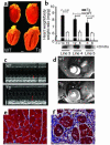
Transgenic expression of Hop in the heart causes cardiac hypertrophy. (a) Wild-type (left) and Hop transgenic (Tg) hearts at 8 weeks of age are shown. Scale bar: 1 mm. (b) Heart weight–to–body weight ratios of Hop transgenic (black bars) and wild-type littermate (white bars) mice between 5 and 10 weeks of age are shown. Hop transgenic hearts are significantly enlarged. (c) M-mode echocardiography demonstrates thickened myocardial walls in transgenic mice (red arrows) and hyperdynamic function with cavity obliteration. (d) EKG-gated cardiac MRI reveals cardiac hypertrophy. Short-axis view images were captured during end diastole showing high contrast between blood (appearing bright) and myocardium. (e) Mason’s trichrome staining of myocardium from wild-type 28-week-old mouse reveals normal cardiac histology. (f) Identical staining technique and magnification of Hop transgenic littermate reveals massive myocyte hypertrophy and significant interstitial fibrosis (blue). (e and f) Scale bars: 50 μm.
Figure 2
Early lethality and cardiac hypertrophy induced by Hop, but not by mutant HopH2. (a) Kaplan-Meier plot revealing reduced survival of Hop transgenic mice (TgHop wt) compared with wild-type and HopH2 transgenic mice (TgHop H2). (b) Cardiac hypertrophy in Hop transgenic mice is progressive. Heart weight–to–body weight ratios were calculated at various ages, as indicated, and expressed as percentage of change compared with wild-type littermates. Values from two independent transgenic lines are pooled and each bar represents the average of 8–16 data points. Error bars represent SEM. *P < 0.05 compared with wild type. (c) Circular dichroism analysis of Hop and HopH2 protein indicates similar conformations and α helicity of wild-type Hop and HopH2 mutant proteins. (θ)MRW, mean residue weight ellipticity. (d). Heart weight–to–body weight ratios of Hop transgenic mice, four independent lines of HopH2 transgenic mice, and Hop knockout mice between 4 and 8 weeks of age. Between 10 and 25 animals were assayed for each condition. No significant difference between transgenic and nontransgenic littermates was determined for HopH2 mice or for _Hop_–/– mice. Western blot analysis of heart tissue using anti-hemagglutinin (HA) antibody revealing expression of transgenic protein is also shown. Line numbers refer to independent transgenic lines. (e) Immunohistochemistry identifies nuclear epitope-tagged Hop protein in transgenic hearts (red). (f) HopH2 is also nuclear localized in transgenic hearts. Scale bars in e and f: 20 μm.
Figure 3
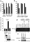
Hop recruits HDAC activity to repress transcription. (a) Cos cells were transfected with a luciferase reporter plasmid including regulatory elements from the SM22α promoter with binding sites for SRF. Myocardin cotransfection induces dramatic activation assigned a value of 100% maximal activation (M). Cotransfection of myocardin and Hop (H) results in inhibition of myocardin-induced activation. The ability of Hop to inhibit myocardin-induced activation is attenuated in a dose-dependent fashion by TSA. (b) Hop inhibitory activity is also blocked by Scriptaid but not by Nullscript. (c) Immunoprecipitation of Hop from transfected Cos cells results in precipitation of HDAC activity. Cells were transfected with vector alone (pcDNA3) or myc-tagged control (pSecTag-PSA), Hop, or HopH2, followed by immunoprecipitation with anti-myc Ab. HDAC activity in the pellet was assayed and reported as arbitrary units (see Methods). Western blot analysis with anti-myc Ab of input material used for immunoprecipitation is shown below. (d) Chromatin immunoprecipitation reveals decrease in acetylated histones after Hop expression. The 10T1/2 cells were transfected as in a, followed by chromatin immunoprecipitation with Ab’s to acetylated histone H4 or H3 and detection of SM22α promoter fragments by PCR in precipitate. (e) Coimmunoprecipitation of HDAC2 and Hop. The 293T cells were transfected with control vector (pcDNA3), Hop, or HopH2, followed by immunoprecipitation and Western blot analysis for HDAC2. A Western blot using anti-HDAC2 Ab of the cell extracts used in each case is shown (Input).
Figure 4
Treatment with HDAC inhibitor prevents cardiac hypertrophy in Hop transgenic mice. (a) HDAC activity was associated with immunoprecipitated transgenic Hop protein derived from hearts of transgenic Hop mice (5–6 weeks of age) when compared with wild-type littermates. (b) Transgenic and wild-type littermates were treated with daily injections of TSA between the ages of 3 and 5 weeks. Heart weight–to–body weight ratios were calculated at 5 weeks. Bars indicate mean of values shown, and each filled circle represents one animal. TSA significantly attenuated cardiac hypertrophy, but did not affect heart weight–to–body weight ratio in nontransgenic mice. (c) Hop transgenic mice were also treated with sodium valproate between 3 and 5 weeks of age, which resulted in attenuation of cardiac enlargement at 5 weeks. (d) Wild-type mice were treated with isoproterenol (ISO) infusion between 3 and 5 weeks of life, with or without daily injection of TSA. Heart weight–to–body weight ratio increased significantly with isoproterenol, and this increase was significantly inhibited by TSA. (e) Model depicting the ability of Hop or other hypertrophic stimuli to recruit class I HDACs resulting in inhibition of antihypertrophic gene programs. Inducers of cardiac hypertrophy are also thought to function by altering calcium homeostasis or mechanical stretch and induce prohypertrophic genes. Class II HDACs can inhibit some prohypertrophic programs. The relative balance of pro- and antihypertrophic gene programs will determine the extent of myocyte hypertrophy.
Comment in
- HATs off to Hop: recruitment of a class I histone deacetylase incriminates a novel transcriptional pathway that opposes cardiac hypertrophy.
Hamamori Y, Schneider MD. Hamamori Y, et al. J Clin Invest. 2003 Sep;112(6):824-6. doi: 10.1172/JCI19834. J Clin Invest. 2003. PMID: 12975465 Free PMC article.
Similar articles
- HATs off to Hop: recruitment of a class I histone deacetylase incriminates a novel transcriptional pathway that opposes cardiac hypertrophy.
Hamamori Y, Schneider MD. Hamamori Y, et al. J Clin Invest. 2003 Sep;112(6):824-6. doi: 10.1172/JCI19834. J Clin Invest. 2003. PMID: 12975465 Free PMC article. - Activation of histone deacetylase 2 by inducible heat shock protein 70 in cardiac hypertrophy.
Kee HJ, Eom GH, Joung H, Shin S, Kim JR, Cho YK, Choe N, Sim BW, Jo D, Jeong MH, Kim KK, Seo JS, Kook H. Kee HJ, et al. Circ Res. 2008 Nov 21;103(11):1259-69. doi: 10.1161/01.RES.0000338570.27156.84. Epub 2008 Oct 10. Circ Res. 2008. PMID: 18849323 - Hop functions downstream of Nkx2.1 and GATA6 to mediate HDAC-dependent negative regulation of pulmonary gene expression.
Yin Z, Gonzales L, Kolla V, Rath N, Zhang Y, Lu MM, Kimura S, Ballard PL, Beers MF, Epstein JA, Morrisey EE. Yin Z, et al. Am J Physiol Lung Cell Mol Physiol. 2006 Aug;291(2):L191-9. doi: 10.1152/ajplung.00385.2005. Epub 2006 Mar 1. Am J Physiol Lung Cell Mol Physiol. 2006. PMID: 16510470 - CAMTA in cardiac hypertrophy.
Schwartz RJ, Schneider MD. Schwartz RJ, et al. Cell. 2006 May 5;125(3):427-9. doi: 10.1016/j.cell.2006.04.015. Cell. 2006. PMID: 16678087 Review. - Roles and post-translational regulation of cardiac class IIa histone deacetylase isoforms.
Weeks KL, Avkiran M. Weeks KL, et al. J Physiol. 2015 Apr 15;593(8):1785-97. doi: 10.1113/jphysiol.2014.282442. Epub 2014 Nov 25. J Physiol. 2015. PMID: 25362149 Free PMC article. Review.
Cited by
- Targeting histone deacetylase in cardiac diseases.
Lu J, Qian S, Sun Z. Lu J, et al. Front Physiol. 2024 Jun 24;15:1405569. doi: 10.3389/fphys.2024.1405569. eCollection 2024. Front Physiol. 2024. PMID: 38983721 Free PMC article. Review. - The Role of TPM3 in Protecting Cardiomyocyte from Hypoxia-Induced Injury via Cytoskeleton Stabilization.
Huang K, Yang W, Shi M, Wang S, Li Y, Xu Z. Huang K, et al. Int J Mol Sci. 2024 Jun 20;25(12):6797. doi: 10.3390/ijms25126797. Int J Mol Sci. 2024. PMID: 38928503 Free PMC article. - Cardiac Transcription Factors and Regulatory Networks.
Grunert M, Dorn C, Rickert-Sperling S. Grunert M, et al. Adv Exp Med Biol. 2024;1441:295-311. doi: 10.1007/978-3-031-44087-8_16. Adv Exp Med Biol. 2024. PMID: 38884718 Review. - Ketones and the Heart: Metabolic Principles and Therapeutic Implications.
Matsuura TR, Puchalska P, Crawford PA, Kelly DP. Matsuura TR, et al. Circ Res. 2023 Mar 31;132(7):882-898. doi: 10.1161/CIRCRESAHA.123.321872. Epub 2023 Mar 30. Circ Res. 2023. PMID: 36996176 Free PMC article. Review. - Combining three independent pathological stressors induces a heart failure with preserved ejection fraction phenotype.
Li Y, Kubo H, Yu D, Yang Y, Johnson JP, Eaton DM, Berretta RM, Foster M, McKinsey TA, Yu J, Elrod JW, Chen X, Houser SR. Li Y, et al. Am J Physiol Heart Circ Physiol. 2023 Apr 1;324(4):H443-H460. doi: 10.1152/ajpheart.00594.2022. Epub 2023 Feb 10. Am J Physiol Heart Circ Physiol. 2023. PMID: 36763506 Free PMC article.
References
- Braunwald, E. 2001. Heart disease: a textbook of cardiovascular medicine. Philadelphia: W.B. Saunders. ch. 13.
- Epstein ND, Davis JS. Sensing stretch is fundamental. Cell. 2003;112:147–150. - PubMed
- McKinsey TA, Olson EN. Cardiac hypertrophy: sorting out the circuitry. Curr. Opin. Genet. Dev. 1999;9:267–274. - PubMed
- Braunwald E. Shattuck lecture—cardiovascular medicine at the turn of the millennium: triumphs, concerns, and opportunities. N. Engl. J. Med. 1997;337:1360–1369. - PubMed
Publication types
MeSH terms
Substances
Grants and funding
- HL-61475/HL/NHLBI NIH HHS/United States
- R21 EB002473-02/EB/NIBIB NIH HHS/United States
- R01 HL061475/HL/NHLBI NIH HHS/United States
- R21 EB002473/EB/NIBIB NIH HHS/United States
- HL-071546/HL/NHLBI NIH HHS/United States
- R01 HL071546/HL/NHLBI NIH HHS/United States
LinkOut - more resources
Full Text Sources
Other Literature Sources
Molecular Biology Databases
Miscellaneous


