Stimulation of medial prefrontal cortex decreases the responsiveness of central amygdala output neurons - PubMed (original) (raw)
Stimulation of medial prefrontal cortex decreases the responsiveness of central amygdala output neurons
Gregory J Quirk et al. J Neurosci. 2003.
Abstract
In extinction of auditory fear conditioning, rats learn that a tone no longer predicts the occurrence of a footshock. Recent lesion and unit recording studies suggest that the medial prefrontal cortex (mPFC) plays an essential role in the inhibition of conditioned fear following extinction. mPFC has robust projections to the amygdala, a structure that is known to mediate the acquisition and expression of conditioned fear. Fear conditioning potentiates the tone responses of neurons in the basolateral amygdala (BLA), which excite neurons in the central nucleus (Ce) of the amygdala. In turn, the Ce projects to the brainstem and hypothalamic areas that mediate fear responses. The present study was undertaken to test the hypothesis that the mPFC inhibits conditioned fear via feedforward inhibition of Ce output neurons. Recording extracellularly from physiologically identified brainstem-projecting Ce neurons, we tested the effect of mPFC prestimulation on Ce responsiveness to synaptic input. In support of our hypothesis, mPFC prestimulation dramatically reduced the responsiveness of Ce output neurons to inputs from the insular cortex and BLA. Thus, our findings support the idea that mPFC gates impulse transmission from the BLA to Ce, perhaps through GABAergic intercalated cells, thereby gating the expression of conditioned fear.
Figures
Figure 1.
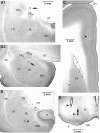
Histological identification of recording (A) and stimulating (B) sites. A, Electrolytic lesions (arrows) made along two different electrode tracks during which we recorded several CeM neurons antidromically responsive to brainstem stimuli. In A1, the lesion marks the site where the first neuron backfired from the brainstem was encountered. In A2, the lesion marks the site where the first neuron antidromically responsive to mPFC stimuli was recorded. B, Tip of stimulating electrodes in the insula (B1), mPFC (B2), and brainstem (B3). The scale bar in A1 is also valid for A2. The scale bar in B1 is also valid for B2. CPU, Striatum; FI, fimbria; H, hippocampus; LA, lateral nucleus of the amygdala; MG, medial geniculate nucleus; OT, optic tract; PAG, periaqueductal gray; RN, red nucleus; rh, rhinal sulcus; SC, superior colliculus; Th, thalamus.
Figure 2.
Examples of antidromic activation of CeM and BL neurons in the rat. A1, Consistent with antidromic activation, brainstem stimulation fired this CeM neuron at a fixed latency (filled circle, 10 overlapped sweeps). A2, A3, Collision test. Collision was observed when spontaneous action potentials occurred within twice the antidromic response latency (A2, 3 overlapped sweeps) but not with longer intervals (A3, 3 overlapped sweeps). B1, Antidromic activation (filled circle) of a BL neuron in response to stimulation of the ipsilateral (ipsi) mPFC. B2, Collision with spikes evoked by suprathreshold stimulation of the contralateral (contra) mPFC. C, Histogram shows the number of neurons (_x_-axis) in either CeM or BL that were antidromically activated by brainstem (right, gray bars) or mPFC (left, white bars) stimuli, respectively, as a function of depth (_y_-axis). Note abrupt transition from antidromic activation of neurons in response to brainstem or mPFC stimuli at the CeM→BL border (arrows on the right), as determined in histological verification of two electrode tracks.
Figure 3.
Inhibition of a CeM output neuron by prestimulation of mPFC in the rat. A, CeM output neuron was synaptically driven by stimulation of the insular cortex (INS). Prestimulation of mPFC reduced the responsiveness of this CeM neuron to insular cortex stimulation (each trace shows 10 overlapping sweeps). Interval between mPFC and insula stimuli is indicated on the left. Inhibition lasted for up to 40 msec in this cell (inset). In the inset, responsiveness was calculated from 20 trials at each ISI. B, Inhibition of antidromic responses of a CeM output neuron. Each trace is the response to a single stimulus. The left column shows responses to brainstem (BS) stimuli applied in isolation. Prestimulation of mPFC (right column) reduced the probability of antidromic invasion of this CeM neuron from 80 to 20% (calculated from 20 trials). The ISI was 20 msec in this case.
Figure 4.
Histological identification of recording (A) and stimulating (B-D) sites. A, Electrolytic lesions made along two different electrode tracks during which we recorded several CeM neurons antidromically responsive to brainstem stimuli. In the top (A1), the lesion marks the site where the first neuron backfired from the brainstem was encountered. In A2, the lesion marks the site where the first neuron antidromically responsive to mPFC stimuli was encountered. B-D, Tip of stimulating electrodes in the BL (B), mPFC (C), and brainstem (D). The scale bar in A1 is also valid for A2. BM, Basomedial nucleus; CA, caudate nucleus; CEL, central lateral nucleus; CEM, central medial nucleus; Cru, cruciate sulcus; EC, external capsule; H, hippocampus; LA, lateral nucleus of the amygdala; OT, optic tract; P, cerebral peduncle; R, red nucleus; SN, substantia nigra; V, ventricle.
Figure 5.
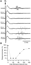
Inhibition of a CeM output neuron by prestimulation of mPFC in the cat. A, BL stimulation activated this CeM output neuron at a latency of ∼7 msec. Prestimulation of ipsilateral mPFC eliminated synaptic responses to BL stimulation. Interval between mPFC and BL stimuli is indicated on the left. B, Graph plotting responsiveness to BL stimuli (_y_-axis) as a function of mPFC-BL ISIs (_x_-axis). Responsiveness was calculated from 20 trials at each ISI. The inhibitory effect of mPFC stimulation lasted ∼120 msec in this neuron.
Figure 6.
Inhibition of BL-evoked field responses in CeM by mPFC prestimulation. Inset shows CeM field response to BL stimulation, both without and with mPFC prestimulation (60 msec ISI). Graph plots BL-evoked field potential amplitude (negative component, _y_-axis) as a function of mPFC-BL ISI (_x_-axis). Note the reduction in negative component amplitude with mPFC prestimulation. mPFC stimuli applied in isolation evoked a positive field potential in CeM (data not shown).
Figure 7.
Scheme depicting the IL-amygdala interactions hypothesized to underlie the phenomenon described in this study. IL inhibits CeM projection neurons via GABAergic ITC cells, thereby reducing CeM responses to inputs from BL or cortex. For clarity, several intramygdala projections have been omitted. BS, Brainstem; IC, internal capsule; OT, optic tract; Pu, putamen; rh, rhinal fissure.
Similar articles
- Prefrontal control of the amygdala.
Likhtik E, Pelletier JG, Paz R, Paré D. Likhtik E, et al. J Neurosci. 2005 Aug 10;25(32):7429-37. doi: 10.1523/JNEUROSCI.2314-05.2005. J Neurosci. 2005. PMID: 16093394 Free PMC article. - Synaptic encoding of fear extinction in mPFC-amygdala circuits.
Cho JH, Deisseroth K, Bolshakov VY. Cho JH, et al. Neuron. 2013 Dec 18;80(6):1491-507. doi: 10.1016/j.neuron.2013.09.025. Epub 2013 Nov 27. Neuron. 2013. PMID: 24290204 Free PMC article. - Prefrontal mechanisms in extinction of conditioned fear.
Quirk GJ, Garcia R, González-Lima F. Quirk GJ, et al. Biol Psychiatry. 2006 Aug 15;60(4):337-43. doi: 10.1016/j.biopsych.2006.03.010. Epub 2006 May 19. Biol Psychiatry. 2006. PMID: 16712801 Review. - New vistas on amygdala networks in conditioned fear.
Paré D, Quirk GJ, Ledoux JE. Paré D, et al. J Neurophysiol. 2004 Jul;92(1):1-9. doi: 10.1152/jn.00153.2004. J Neurophysiol. 2004. PMID: 15212433 Review.
Cited by
- Attachment models affect brain responses in areas related to emotions and empathy in nulliparous women.
Lenzi D, Trentini C, Pantano P, Macaluso E, Lenzi GL, Ammaniti M. Lenzi D, et al. Hum Brain Mapp. 2013 Jun;34(6):1399-414. doi: 10.1002/hbm.21520. Epub 2012 Feb 22. Hum Brain Mapp. 2013. PMID: 22359374 Free PMC article. - Chronic treatment with corticosterone increases the number of tyrosine hydroxylase-expressing cells within specific nuclei of the brainstem reticular formation.
Busceti CL, Bucci D, Scioli M, Di Pietro P, Nicoletti F, Puglisi-Allegra S, Ferrucci M, Fornai F. Busceti CL, et al. Front Neuroanat. 2022 Oct 28;16:976714. doi: 10.3389/fnana.2022.976714. eCollection 2022. Front Neuroanat. 2022. PMID: 36387998 Free PMC article. - Adaptive contextualization: A new role for the default mode network in affective learning.
Marstaller L, Burianová H, Reutens DC. Marstaller L, et al. Hum Brain Mapp. 2017 Feb;38(2):1082-1091. doi: 10.1002/hbm.23442. Epub 2016 Oct 21. Hum Brain Mapp. 2017. PMID: 27767246 Free PMC article. - The nature of individual differences in inhibited temperament and risk for psychiatric disease: A review and meta-analysis.
Clauss JA, Avery SN, Blackford JU. Clauss JA, et al. Prog Neurobiol. 2015 Apr;127-128:23-45. doi: 10.1016/j.pneurobio.2015.03.001. Epub 2015 Mar 14. Prog Neurobiol. 2015. PMID: 25784645 Free PMC article. Review. - Network model of fear extinction and renewal functional pathways.
Bruchey AK, Shumake J, Gonzalez-Lima F. Bruchey AK, et al. Neuroscience. 2007 Mar 16;145(2):423-37. doi: 10.1016/j.neuroscience.2006.12.014. Epub 2006 Dec 16. Neuroscience. 2007. PMID: 17257766 Free PMC article.
References
- al Maskati HA, Zbrozyna AW ( 1989) Stimulation in prefrontal cortex area inhibits cardiovascular and motor components of the defence reaction in rats. J Auton Nerv Syst 28: 117-125. - PubMed
- Bellgowan PS, Helmstetter FJ ( 1996) Neural systems for the expression of hypoalgesia during nonassociative fear. Behav Neurosci 110: 727-736. - PubMed
- Blair HT, Schafe GE, Bauer EP, Rodrigues SM, LeDoux JE ( 2001) Synaptic plasticity in the lateral amygdala: a cellular hypothesis of fear conditioning. Learn Mem 8: 229-242. - PubMed
- Bouton ME, King DA ( 1983) Contextual control of the extinction of conditioned fear: tests for the associative value of the context. J Exp Psychol Anim Behav Process 9: 248-265. - PubMed
Publication types
MeSH terms
LinkOut - more resources
Full Text Sources
Other Literature Sources


