Reverse genetics with a full-length infectious cDNA of severe acute respiratory syndrome coronavirus - PubMed (original) (raw)
Reverse genetics with a full-length infectious cDNA of severe acute respiratory syndrome coronavirus
Boyd Yount et al. Proc Natl Acad Sci U S A. 2003.
Abstract
A previously undescribed coronavirus (CoV) is the etiologic agent responsible for severe acute respiratory syndrome (SARS). Using a panel of contiguous cDNAs that span the entire genome, we have assembled a full-length cDNA of the SARS-CoV Urbani strain, and have rescued molecularly cloned SARS viruses (infectious clone SARS-CoV) that contained the expected marker mutations inserted into the component clones. Recombinant viruses replicated as efficiently as WT virus and both were inhibited by treatment with the cysteine proteinase inhibitor (2S,3S)-transepoxysuccinyl-L-leucylamido-3-methylbutane ethyl ester. In addition, subgenomic transcripts were initiated from the consensus sequence ACGAAC in both the WT and infectious clone SARS-CoV. Availability of a SARS-CoV full-length cDNA provides a template for manipulation of the viral genome, allowing for the rapid and rational development and testing of candidate vaccines and therapeutics against this important human pathogen.
Figures
Fig. 1.
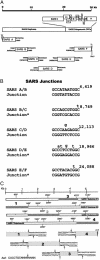
Assembly of a full-length SARS-CoV cDNA. (A) Structure of the SARS-CoV genome (8, 9). The SARS Urbani ≈29,727-bp genome contains 10 or more ORFs, which are expressed from full-length and subgenomic-length mRNAs (8). (B) Several _Bgl_I-interconnecting junctions were inserted between the component clones to allow assembly of a full-length cDNA. Lowercase letters represent WT sequence and numbers represent nucleotide positions in genome. (C) “No see'm” Aar I repair of SARS sibling clones. Asterisks represent sites of mutation. Numbers in bold represent portions of sibling clones that were assembled into a consensus SARS F subclone.
Fig. 2.
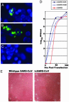
SARS-CoV full-length transcripts are infectious. Full-length transcripts, in the presence or absence of SARS N transcripts, were electroporated into BHK cells and overlaid with susceptible VeroE6 cells. A portion of the cells was examined by fluorescent Ab staining. (A) Cultures transfected with SARS-CoV full-length transcripts. (B) SARS full-length transcripts plus N transcripts. (C) Uninfected control. (D) Virus growth was determined for the first 96 h after transfection by plaque assay; icSARS transcripts alone (▪) and icSARS plus N transcripts, trials 1 (⋄) and 2 (▵). (E) icSARS-CoV and WT SARS-CoV plaques in Vero E6 cells.
Fig. 3.
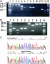
icSARS-CoV marker mutations. cDNAs spanning the different _Bgl_I junctions were amplified from WT and icSARS-CoV-infected cells. The fragments were purified, and a portion was digested with _Bgl_I and separated in 1.2% agarose gels. (A) icSARS-CoV (lanes 2, 3, 6, 7, 10, and 11) and WT SARS-CoV (lanes 4, 5, 8, 9, 12, and 13). Lanes 2–5, a 1,668-nt amplicon (nucleotide positions 1007–2675) digested with _Bgl_I (lanes 3 and 5); lanes 6–9, a 799-nt amplicon spanning the SARS-CoV B/C junction (positions 8381–9180) digested with _Bgl_I (lanes 7 and 9); lanes 10–13, a 544-nt amplicon (positions 11,721–12,265) spanning the SARS-CoV C/D junction digested with _Bgl_I (lanes 11 and 13). Lanes 1 and 14, a 1-kb ladder. (B) WT SARS (lanes 4, 5, 8, and 9); icSARS-CoV (lanes 2, 3, 6, and 7). Lanes 2–5, a 652-nt amplicon spanning the SARS-CoV D/E junction digested with _Bgl_I (lanes 3 and 5); lanes 6–9, a 1,594-nt amplicon (positions 23,665–25,259) spanning the SARS-CoV E/F junction digested with _Bgl_I (lanes 7 and 9). Lanes 1 and 10, a 1-kb ladder. (C) The 1,594-nt SARS E/F junction-containing amplicon was subcloned and sequenced, demonstrating the mutations introduced within the icSARS-CoV.
Fig. 4.
Phenotype comparisons between WT and icSARS-CoV. Cultures of cells were infected with an multiplicity of infection of 0.1. At 1 h, cultures were treated with E64-d at a concentration of 500 μg/ml, and virus titers were determined by plaque assay in VeroE6 cells (C). (A) Growth of Urbani (▪), icSARS (□), and the Tor-2 (▴) and Tor-7 (▵) CoVs. (B) Leader-containing transcripts encoding various icSARS-CoV subgenomic ORFs. *, predicted ORFs classified by using standard CoV nomenclature (11). (C) Growth of icSARS and Urbani SARS-CoV in the presence or absence of E64-d. Hatched bars, 24-h titers; black bars, 48-h titers. (D) Urbani SARS-CoV-infected cells at 72 h after infection.
Similar articles
- [Construction of genomic full-length cDNA of SARS coronavirus BJ01 strain and identification of its biological characteristics].
Han JF, Jiang T, Chen SP, Yu M, Qin ED, Zhao Z, Li XF, Qin CF, Deng YQ, Zhao H, Zha HL, Li XY. Han JF, et al. Wei Sheng Wu Xue Bao. 2006 Dec;46(6):922-7. Wei Sheng Wu Xue Bao. 2006. PMID: 17302155 Chinese. - Reverse genetics with a full-length infectious cDNA of the Middle East respiratory syndrome coronavirus.
Scobey T, Yount BL, Sims AC, Donaldson EF, Agnihothram SS, Menachery VD, Graham RL, Swanstrom J, Bove PF, Kim JD, Grego S, Randell SH, Baric RS. Scobey T, et al. Proc Natl Acad Sci U S A. 2013 Oct 1;110(40):16157-62. doi: 10.1073/pnas.1311542110. Epub 2013 Sep 16. Proc Natl Acad Sci U S A. 2013. PMID: 24043791 Free PMC article. - Construction of a severe acute respiratory syndrome coronavirus infectious cDNA clone and a replicon to study coronavirus RNA synthesis.
Almazán F, Dediego ML, Galán C, Escors D, Alvarez E, Ortego J, Sola I, Zuñiga S, Alonso S, Moreno JL, Nogales A, Capiscol C, Enjuanes L. Almazán F, et al. J Virol. 2006 Nov;80(21):10900-6. doi: 10.1128/JVI.00385-06. Epub 2006 Aug 23. J Virol. 2006. PMID: 16928748 Free PMC article. - Development of mouse hepatitis virus and SARS-CoV infectious cDNA constructs.
Baric RS, Sims AC. Baric RS, et al. Curr Top Microbiol Immunol. 2005;287:229-52. doi: 10.1007/3-540-26765-4_8. Curr Top Microbiol Immunol. 2005. PMID: 15609514 Free PMC article. Review. - Understanding the accessory viral proteins unique to the severe acute respiratory syndrome (SARS) coronavirus.
Tan YJ, Lim SG, Hong W. Tan YJ, et al. Antiviral Res. 2006 Nov;72(2):78-88. doi: 10.1016/j.antiviral.2006.05.010. Epub 2006 Jun 6. Antiviral Res. 2006. PMID: 16820226 Free PMC article. Review.
Cited by
- Establishment of a reverse genetics system for SARS-CoV-2 using circular polymerase extension reaction.
Torii S, Ono C, Suzuki R, Morioka Y, Anzai I, Fauzyah Y, Maeda Y, Kamitani W, Fukuhara T, Matsuura Y. Torii S, et al. Cell Rep. 2021 Apr 20;35(3):109014. doi: 10.1016/j.celrep.2021.109014. Epub 2021 Apr 1. Cell Rep. 2021. PMID: 33838744 Free PMC article. - A Perspective on COVID-19 Management.
Pavelić K, Kraljević Pavelić S, Brix B, Goswami N. Pavelić K, et al. J Clin Med. 2021 Apr 9;10(8):1586. doi: 10.3390/jcm10081586. J Clin Med. 2021. PMID: 33918624 Free PMC article. Review. - Evidence supporting a zoonotic origin of human coronavirus strain NL63.
Huynh J, Li S, Yount B, Smith A, Sturges L, Olsen JC, Nagel J, Johnson JB, Agnihothram S, Gates JE, Frieman MB, Baric RS, Donaldson EF. Huynh J, et al. J Virol. 2012 Dec;86(23):12816-25. doi: 10.1128/JVI.00906-12. Epub 2012 Sep 19. J Virol. 2012. PMID: 22993147 Free PMC article. - Partial and full PCR-based reverse genetics strategy for influenza viruses.
Chen H, Ye J, Xu K, Angel M, Shao H, Ferrero A, Sutton T, Perez DR. Chen H, et al. PLoS One. 2012;7(9):e46378. doi: 10.1371/journal.pone.0046378. Epub 2012 Sep 28. PLoS One. 2012. PMID: 23029501 Free PMC article. - Increased antibody affinity confers broad in vitro protection against escape mutants of severe acute respiratory syndrome coronavirus.
Rani M, Bolles M, Donaldson EF, Van Blarcom T, Baric R, Iverson B, Georgiou G. Rani M, et al. J Virol. 2012 Sep;86(17):9113-21. doi: 10.1128/JVI.00233-12. Epub 2012 Jun 13. J Virol. 2012. PMID: 22696652 Free PMC article.
References
- Tsang, K. W., Ho, P. L., Ooi, G. C., Yee, W. K., Wang, T., Chan-Yeung, M., Lam, W. K., Seto, W. H., Yam, L. Y., Cheung, T. M., et al. (2003) N. Engl. J. Med. 348 1977-1985. - PubMed
- Lee, N., Hui, D., Wu, A., Chan, P., Cameron, P., Joynt, G. M., Ahuja, A., Yung, M. Y., Leung, C. B, To, K. F., et al. (2003) N. Engl. J. Med. 348 1986-1994. - PubMed
- Poutanen, S. M., Low, D. E., Henry, B., Finkelstein, S., Rose, D., Green, K., Tellier, R., Draker, R., Adachi, D., Ayers, M., et al. (2003) N. Engl. J. Med. 348 1995-2005. - PubMed
Publication types
MeSH terms
Substances
Grants and funding
- GM 63228/GM/NIGMS NIH HHS/United States
- R01 AI026603/AI/NIAID NIH HHS/United States
- AI 26603/AI/NIAID NIH HHS/United States
- AI 23946/AI/NIAID NIH HHS/United States
- R01 AI023946/AI/NIAID NIH HHS/United States
- R01 GM063228/GM/NIGMS NIH HHS/United States
LinkOut - more resources
Full Text Sources
Other Literature Sources
Miscellaneous


