An increased high-mobility group A2 expression level is associated with malignant phenotype in pancreatic exocrine tissue - PubMed (original) (raw)
An increased high-mobility group A2 expression level is associated with malignant phenotype in pancreatic exocrine tissue
N Abe et al. Br J Cancer. 2003.
Abstract
The altered form of the high-mobility group A2 (HMGA2) gene is somehow related to the generation of human benign and malignant tumours of mesenchymal origin. However, only a few data on the expression of HMGA2 in malignant tumour originating from epithelial tissue are available. In this study, we examined the HMGA2 expression level in pancreatic carcinoma, and investigated whether alterations in the HMGA2 expression level are associated with a malignant phenotype in pancreatic tissue. High-mobility group A2 mRNA and protein expression was determined in eight surgically resected specimens of non-neoplastic tissue (six specimens of normal pancreatic tissue and two of chronic pancreatitis tissue) and 27 pancreatic carcinomas by highly sensitive reverse transcriptase-polymerase chain reaction (RT-PCR) techniques and immunohistochemical staining, respectively. Reverse transcriptase-polymerase chain reaction analysis revealed the expression of the HMGA2 gene in non-neoplastic pancreatic tissue, although its expression level was significantly lower than that in carcinoma. Immunohistochemical analysis indicated that the presence of the HMGA2 gene in non-neoplastic pancreatic tissue observed in RT-PCR reflects its abundant expression in islet cells, together with its focal expression in duct epithelial cells. Intense and multifocal or diffuse HMGA2 immunoreactivity was noted in all the pancreatic carcinoma examined. A strong correlation between HMGA2 overexpression and the diagnosis of carcinoma was statistically verified. Based on these findings, we propose that an increased expression level of the HMGA2 protein is closely associated with the malignant phenotype in the pancreatic exocrine system, and accordingly, HMGA2 could serve as a potential diagnostic molecular marker for distinguishing pancreatic malignant cells from non-neoplastic pancreatic exocrine cells.
Figures
Figure 1
Reverse transcriptase–polymerase chain reaction products of HMGA2 after gel electrophoresis and ethidium bromide staining. Results show specific 220-bp bands. DL, DNA molecular weight marker; lane 1, positive control (hepatoma cell line HEP3B, which is known to express high level of HMGA2); lane 2, normal pancreas; lane 3, chronic pancreatitis; lane 4–11, pancreatic carcinomas.
Figure 2
Immunohistochemical demonstration of the HMGA2 protein expression in pancreatic cancer cell lines. (A) AsPC-1 (Mayer's haematoxylin; original magnification × 200). (B) PANC-I (Mayer's haematoxylin; original magnification × 200). (C) MIA PaCa-2 (Mayer's haematoxylin; original magnification × 100). (D) BxPC-3 (Mayer's haematoxylin; original magnification × 200). Intense multifocal or diffuse HMGA2 nuclear immunoreactivity (brown colour) was characteristically observed in cancer cells.
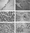
Figure 3
Immunohistochemical demonstration of the HMGA2 protein expression in surgically resected specimens of non-neoplastic pancreatic tissues and pancreatic carcinomas. (A) Non-neoplastic epithelial cells of the main pancreatic duct. A small proportion of duct epithelial cells show HMGA2 immunoreactivity (arrows). (Mayer's haematoxylin; original magnification × 200). (B) Epithelial cells of branch pancreatic duct and islets in chronic pancreatitis tissue. Islet cells showed intense and diffuse HMGA2 immunoreactivity (arrows), while epithelial cells of the branch pancreatic duct did not exhibit any detectable HMGA2 immunoreactivity (arrowhead) (Mayer's haematoxylin; original magnification × 100). (C) Primary pancreatic carcinoma exhibiting well-differentiated tubular adenocarcinoma (Mayer's haematoxylin; original magnification × 200). (D) Primary pancreatic carcinoma exhibiting adenosquamous carcinoma (Mayer's haematoxylin; original magnification × 200). (E) Metastatic lesion in the liver (Mayer's haematoxylin; original magnification × 200). Intense and multifocal or diffuse HMGA2 immunoreactivity was noted in all the pancreatic carcinomas (C–E). (F) Section including both carcinoma cells and islet cells (Mayer's haematoxylin; original magnification × 200). Islet cells showed intense and diffuse HMGA2 immunoreactivity (arrows), which was almost equivalent to that observed in carcinoma cells (arrowheads).
Similar articles
- HMGA2 protein expression correlates with lymph node metastasis and increased tumor grade in pancreatic ductal adenocarcinoma.
Hristov AC, Cope L, Reyes MD, Singh M, Iacobuzio-Donahue C, Maitra A, Resar LM. Hristov AC, et al. Mod Pathol. 2009 Jan;22(1):43-9. doi: 10.1038/modpathol.2008.140. Epub 2008 Aug 29. Mod Pathol. 2009. PMID: 18843278 Free PMC article. - HMGA1 and HMGA2 protein expression correlates with advanced tumour grade and lymph node metastasis in pancreatic adenocarcinoma.
Piscuoglio S, Zlobec I, Pallante P, Sepe R, Esposito F, Zimmermann A, Diamantis I, Terracciano L, Fusco A, Karamitopoulou E. Piscuoglio S, et al. Histopathology. 2012 Feb;60(3):397-404. doi: 10.1111/j.1365-2559.2011.04121.x. Histopathology. 2012. PMID: 22276603 - Prognostic significance of high mobility group A2 (HMGA2) in pancreatic ductal adenocarcinoma: malignant functions of cytoplasmic HMGA2 expression.
Gundlach JP, Hauser C, Schlegel FM, Willms A, Halske C, Röder C, Krüger S, Röcken C, Becker T, Kalthoff H, Trauzold A. Gundlach JP, et al. J Cancer Res Clin Oncol. 2021 Nov;147(11):3313-3324. doi: 10.1007/s00432-021-03745-w. Epub 2021 Jul 24. J Cancer Res Clin Oncol. 2021. PMID: 34302528 Free PMC article. - Pancreatic duct cell carcinomas express high levels of high mobility group I(Y) proteins.
Abe N, Watanabe T, Masaki T, Mori T, Sugiyama M, Uchimura H, Fujioka Y, Chiappetta G, Fusco A, Atomi Y. Abe N, et al. Cancer Res. 2000 Jun 15;60(12):3117-22. Cancer Res. 2000. PMID: 10866296 - The MUC gene family: their role in diagnosis and early detection of pancreatic cancer.
Ringel J, Löhr M. Ringel J, et al. Mol Cancer. 2003 Jan 7;2:9. doi: 10.1186/1476-4598-2-9. Mol Cancer. 2003. PMID: 12556240 Free PMC article. Review.
Cited by
- Significance of High-Mobility Group A Protein 2 Expression in Pancreatic Ductal Adenocarcinoma and Ampullary Adenocarcinoma.
Oflas D, Canaz F, Özer İ, Demir L, Çolak E. Oflas D, et al. Turk J Gastroenterol. 2023 Oct;34(10):1014-1024. doi: 10.5152/tjg.2023.22881. Turk J Gastroenterol. 2023. PMID: 37787719 Free PMC article. - LncRNA HOTAIR influences cell growth, migration, invasion, and apoptosis via the miR-20a-5p/HMGA2 axis in breast cancer.
Zhao W, Geng D, Li S, Chen Z, Sun M. Zhao W, et al. Cancer Med. 2018 Mar;7(3):842-855. doi: 10.1002/cam4.1353. Epub 2018 Feb 23. Cancer Med. 2018. PMID: 29473328 Free PMC article. Retracted. - Three-dimensional collagen I promotes gemcitabine resistance in pancreatic cancer through MT1-MMP-mediated expression of HMGA2.
Dangi-Garimella S, Krantz SB, Barron MR, Shields MA, Heiferman MJ, Grippo PJ, Bentrem DJ, Munshi HG. Dangi-Garimella S, et al. Cancer Res. 2011 Feb 1;71(3):1019-28. doi: 10.1158/0008-5472.CAN-10-1855. Epub 2010 Dec 8. Cancer Res. 2011. PMID: 21148071 Free PMC article. - Small interfering-high mobility group A2 attenuates epithelial-mesenchymal transition in thymic cancer cells via the Wnt/β-catenin pathway.
Tan S, Chen J. Tan S, et al. Oncol Lett. 2021 Aug;22(2):586. doi: 10.3892/ol.2021.12847. Epub 2021 Jun 3. Oncol Lett. 2021. PMID: 34122637 Free PMC article. - Multi-omics data integration and modeling unravels new mechanisms for pancreatic cancer and improves prognostic prediction.
Fraunhoffer NA, Abuelafia AM, Bigonnet M, Gayet O, Roques J, Nicolle R, Lomberk G, Urrutia R, Dusetti N, Iovanna J. Fraunhoffer NA, et al. NPJ Precis Oncol. 2022 Aug 17;6(1):57. doi: 10.1038/s41698-022-00299-z. NPJ Precis Oncol. 2022. PMID: 35978026 Free PMC article.
References
- Abe N, Watanabe T, Izumisato Y, Masaki T, Mori T, Sugiyama M, Chiappetta G, Fusco A, Fujioka Y, Atomi Y (2002) Diagnostic significance of high mobility group I(Y) protein expression in intraductal papillary mucinous tumors of the pancreas. Pancreas 25: 149–153 - PubMed
- Abe N, Watanabe T, Masaki T, Mori T, Sugiyama M, Uchimura H, Fujioka Y, Chiappetta G, Fusco A, Atomi Y (2000) Pancreatic duct cell carcinomas express high levels of high mobility group I(Y) (HMGI(Y)) proteins. Cancer Res 60: 3117–3122 - PubMed
- Abe N, Watanabe T, Sugiyama M, Uchimura H, Chiappetta G, Fusco A, Atomi Y (1999) Determination of high mobility group I(Y) expression level in colorectal neoplasias: a potential diagnostic marker. Cancer Res 59: 1169–1174 - PubMed
- Arlotta P, Tai AK, Manfioletti G, Clifford C, Jay G, Ono SJ (2000) Transgenic mice expressing a truncated form of the high mobility group I–C protein develop adiposity and an abnormally high prevalence of lipomas. J Biol Chem 275: 14394–14400 - PubMed
- Ashar HR, Fejzo MS, Tkachenko A, Zhou X, Fletcher JA, Weremowicz S, Morton CC, Chada K (1995) Disruption of the architectural factor HMGI-C: DNA-binding AT hook motifs fused in lipomas to distinct transcriptional regulatory domains. Cell 82: 57–65 - PubMed
Publication types
MeSH terms
Substances
LinkOut - more resources
Full Text Sources
Other Literature Sources
Medical


