Microvascularization of the human digit as studied by corrosion casting - PubMed (original) (raw)
Microvascularization of the human digit as studied by corrosion casting
S Sangiorgi et al. J Anat. 2004 Feb.
Abstract
The aim of this study was to describe microcirculation in the human digit, focusing on the vascular patterns of its cutaneous and subcutaneous areas. We injected a functional supranumerary human thumb (Wassel type IV) with a low-viscosity acrylic resin through its digital artery. The tissues around the vessels were then digested in hot alkali and the resulting casts treated for scanning electron microscopy. We concentrated on six different areas: the palmar and dorsal side of the skin, the eponychium, the perionychium, the nail bed and the nail root. On the palmar side, many vascular villi were evident: these capillaries followed the arrangement of the fingerprint lines, whereas on the dorsal side they were scattered irregularly inside the dermal papillae. In the hypodermal layer of the palmar area, vascular supports of sweat glands and many arteriovenous anastomoses were visible, along with glomerular-shaped vessels involved in thermic regulation and tactile function. In the eponychium and perionychium, the vascular villi followed the direction of nail growth. In the face of the eponychium in contact with the nail, a wide-mesh net of capillaries was evident. In the nail bed, the vessels were arranged in many longitudinal trabeculae parallel to the major axis of the digit. In the root of the nail, we found many columnar vessels characterized by multiple angiogenic buttons on their surface.
Figures
Fig. 1
Schematic representation of the digit in all six areas investigated: palmar (A) and dorsal (B) side of digital skin, eponychium (C), perionychium (D), nail bed (E) and nail root (F): the arrows connect each area to the studied region.
Fig. 2
Palmar side. In transverse section we can distinguish four vascular layers: (1) the papillary layer, (2) the subpapillary layer, (3) the reticular layer and (4) the hypodermal layer: note the glomerular structures supplying sweat glands and thermoreceptorial systems. We can also distinguish the arterial (A) from venular (V) vessels.
Fig. 3
Palmar side. The digit is seen during the maceration process on the 2nd day of KOH immersion. It already shows the regular linear disposition of vessels along the fingerprints (A); (B) superimposition of an SEM image of the same area, revealing perfect correspondence.
Fig. 4
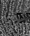
Palmar and dorsal side. In the palmar side we can observe two rows of vessels following the patterning the fingerprint. (A) The villi that arise from these vessels are 100 µm high and have a dextrogyrate rotation: the distance between each is about 70 µm (B). On the dorsal side the vessels do not follow any pattern (C).
Fig. 5
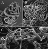
Palmar side. In the hypodermal layer of the palmar side, we can observe many glomeruloid vascular structures: the vessels supplying the sweat glands (A), the capillaries surrounding thermoreceptorial corpuscles (B) and some glomic bodies or arteriovenous anastomoses (C). In the glomic body, we can distinguish the artery (a) from the vein (v) by looking at the nuclear impressions left on the cast by the endothelial cells (thin arrow).
Fig. 6
Eponychium. The angle formed between the axis of the vascular villi on the dorsal side of the skin and the major axis of the digit progressively diminishes towards the eponychium (thin arrow) – nail (thick arrow) border.
Fig. 7
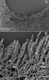
Eponychium. At lower magnification we can clearly see the convexity of the face in contact with the nail (A). At high magnification we can distinguish a regular vascular net that gives rise to the first long vascular villi of the eponychium (B). We can distinguish the artery (a) from the vein (v) by their different morphological conformation.
Fig. 8
Perionychium. The passage from the nail bed to the peryonichium is made up of an interlaced net of thin vessels, where the pressure of the nail on the tissue is higher, going from the trabecular area of the nail bed to the vascular villi of the skin region.
Fig. 9
Nail bed. Two different patterns of disposition of vascular trabeculae: the vessels begin to form a number of loops, longitudinally orientated along the major axis of the nail to the passage to the eponychium (A). The vascular loops have a gradual coronal inclination in the middle of the nail bed (B).
Fig. 10
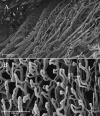
Nail root. The vascular architecture is composed of an area with helicoidal-shaped vessels that gradually turn into longitudinal vessels, giving rise to the trabeculae of the nail bed (A). These vessels are spiral-shaped when they originate, and gradually straighten (B).
Fig. 11
Nail root. On the helicoidal vessels found in the nail root a number of angiogenic buttons are visible (A). At high magnification, we can distinguish different patterns of conformation: formation of collaterals (B) and dome-shaped elevations (C).
Similar articles
- Blood vessels in the tongue of the kitten: scanning electron microscopy of microcorrosion casts.
Jasiński A, Miodoński A. Jasiński A, et al. Anat Embryol (Berl). 1979 Apr 6;155(3):347-53. doi: 10.1007/BF00317647. Anat Embryol (Berl). 1979. PMID: 453549 - The cutaneous microvascular architecture of human diabetic toe studied by corrosion casting and scanning electron microscopy analysis.
Sangiorgi S, Manelli A, Reguzzoni M, Ronga M, Protasoni M, Dell'Orbo C. Sangiorgi S, et al. Anat Rec (Hoboken). 2010 Oct;293(10):1639-45. doi: 10.1002/ar.21168. Epub 2010 Aug 4. Anat Rec (Hoboken). 2010. PMID: 20687174 - A new method for the joint visualization of vascular structures and connective tissues: corrosion casting and 1 N NaOH maceration.
Sangiorgi S, Manelli A, Dell'Orbo C, Congiu T. Sangiorgi S, et al. Microsc Res Tech. 2006 Nov;69(11):919-23. doi: 10.1002/jemt.20366. Microsc Res Tech. 2006. PMID: 16921528 - Scanning electron microscopy of corrosion casting in medicine.
Konerding MA. Konerding MA. Scanning Microsc. 1991 Sep;5(3):851-65. Scanning Microsc. 1991. PMID: 1725561 Review. - Evaluation of several methods used in anatomical investigations of the blood and lymphatic vessels.
Jedrzejewski KS, Cendrowska I, Okraszewska E. Jedrzejewski KS, et al. Folia Morphol (Warsz). 2002;61(2):63-9. Folia Morphol (Warsz). 2002. PMID: 12164052 Review.
Cited by
- Synchrotron phase-contrast imaging applied to the anatomical study of the hand and its vascularization.
Solé Cruz E, Mercier A, Suuronen JP, Chaffanjon P, Brun E, Bellier A. Solé Cruz E, et al. J Anat. 2021 Aug;239(2):536-543. doi: 10.1111/joa.13427. Epub 2021 Mar 8. J Anat. 2021. PMID: 33686643 Free PMC article. - The dermal arteries of the human thumb pad.
Geyer SH, Nöhammer MM, Tinhofer IE, Weninger WJ. Geyer SH, et al. J Anat. 2013 Dec;223(6):603-9. doi: 10.1111/joa.12113. Epub 2013 Sep 20. J Anat. 2013. PMID: 24205910 Free PMC article. - The reason for red streaks on dermoscopy in the distal part of a subungual hemorrhage.
Sato T, Tanaka M. Sato T, et al. Dermatol Pract Concept. 2014 Apr 30;4(2):83-5. doi: 10.5826/dpc.0402a18. eCollection 2014 Apr. Dermatol Pract Concept. 2014. PMID: 24855582 Free PMC article. No abstract available. - Capturing the vital vascular fingerprint with optical coherence tomography.
Liu G, Chen Z. Liu G, et al. Appl Opt. 2013 Aug 1;52(22):5473-7. doi: 10.1364/AO.52.005473. Appl Opt. 2013. PMID: 23913068 Free PMC article. - The dermal arteries in the cutaneous angiosome of the descending genicular artery.
Tinhofer IE, Zaussinger M, Geyer SH, Meng S, Kamolz LP, Tzou CH, Weninger WJ. Tinhofer IE, et al. J Anat. 2018 Jun;232(6):979-986. doi: 10.1111/joa.12792. Epub 2018 Feb 14. J Anat. 2018. PMID: 29441575 Free PMC article.
References
- Backhouse KM. The blood supply of the arm and hand. In: Saunders WB, editor. The Hand. Philadelphia: Tubiana R; 1981. pp. 297–309. <10.1111/j.1469-7580.2004.00251.x>. - DOI
- Blanka S, Alter M. Dermatoglyphics in Medical Disorders. New York: Springer-Verlag; 1976. <10.1111/j.1469-7580.2004.00251.x>. - DOI
MeSH terms
LinkOut - more resources
Full Text Sources
Other Literature Sources


