Properties of replication-competent vesicular stomatitis virus vectors expressing glycoproteins of filoviruses and arenaviruses - PubMed (original) (raw)
Properties of replication-competent vesicular stomatitis virus vectors expressing glycoproteins of filoviruses and arenaviruses
Michael Garbutt et al. J Virol. 2004 May.
Abstract
Replication-competent recombinant vesicular stomatitis viruses (rVSVs) expressing the type I transmembrane glycoproteins and selected soluble glycoproteins of several viral hemorrhagic fever agents (Marburg virus, Ebola virus, and Lassa virus) were generated and characterized. All recombinant viruses exhibited rhabdovirus morphology and replicated cytolytically in tissue culture. Unlike the rVSVs with an additional transcription unit expressing the soluble glycoproteins, the viruses carrying the foreign transmembrane glycoproteins in replacement of the VSV glycoprotein were slightly attenuated in growth. Biosynthesis and processing of the foreign glycoproteins were authentic, and the cell tropism was defined by the transmembrane glycoprotein. None of the rVSVs displayed pathogenic potential in animals. The rVSV expressing the Zaire Ebola virus transmembrane glycoprotein mediated protection in mice against a lethal Zaire Ebola virus challenge. Our data suggest that the recombinant VSV can be used to study the role of the viral glycoproteins in virus replication, immune response, and pathogenesis.
Figures
FIG. 1.
Schematic drawing of the infectious clone system for VSV, Indiana serotype. BSR-T7 cells were cotransfected with a plasmid containing the VSV genome (VSVXN2 or VSVXN2ΔG) and plasmids expressing the VSV nucleoprotein (pBS-VSV N), phosphoprotein (pBS-VSV P), or polymerase (pBS-VSV L). Transcription of all plasmids is under the control of the bacteriophage T7 RNA promoter. For this study, the glycoproteins of Zaire Ebola virus (ZEBOV GP), Marburg virus (MARV GP), and Lassa virus (LASV GPC) were inserted between the VSV matrix and polymerase (L) genes by using plasmid VSVXN2ΔG (A). In addition, Zaire Ebola virus sGP and Marburg virus GP1 were inserted as an additional gene into vector VSVXN2 (B).
FIG. 2.
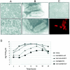
Characterization of rescued rVSVs. Rescued rVSVs were used to infect VeroE6 cells at an MOI of 0.1 PFU/cell. (A) Cytopathogenic effect of infected VeroE6 cells is shown by phase-contrast microscopy 24 h postinfection with VSVΔG/MARVGP (lower panel) in comparison with a mock-infected culture (upper panels). (B) Particle morphology. Electron micrographs show wild-type VSV (upper panel) and VSVΔG/MARVGP (lower panel). (C) Immunofluorescence staining of VeroE6 cells infected with VSVΔG/MARVGP with the GP-specific monoclonal antibody 5EII (dilution, 1:1,000) (lower panel). The upper panel shows the same cells in bright-field microscopy. (D) Growth curves. VeroE6 cells were infected with wild-type VSV (VSVwt), VSV/ZEBOVsGP, VSV/MARVGP1, VSVΔG/ZEBOVGP, VSVΔG/LASVGP, or VSVΔG/MARVGP at an MOI of 10. Supernatants were collected at the indicated times and titrated by defining the TCID50.
FIG. 3.
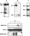
Biosynthesis of the foreign glycoproteins expressed after infection with rVSVs. VeroE6 cells were infected with rVSVs at an MOI of 10. (A) For cells infected with VSVΔG/MARVGP, proteins were pulse labeled at 24 h postinfection for 30 min with 20 μCi of [35S]cysteine per ml and chased for 240 min. GP-specific proteins were immunoprecipitated from cell lysates with mouse anti-Marburg virus GP immunoglobulin (II9G4) (dilution, 1:800) and analyzed on SDS-10% PAGE under reducing conditions. The presence of decRVKR (25 μM) during labeling and chase abolished cleavage of pre-GP (lane 2). (B) For cells infected with VSVΔG/EBOVGP, cells were lysed 24 h postinfection and analyzed by Western blotting with a GP1-specific antibody at a dilution of 1:4,000 (lane 1) and GP2-specific rabbit antiserum at a dilution of 1:2,000 (lane 2). (C) For cells infected with VSVΔG/LASVGPC, cells were lysed 24 h postinfection and analyzed by Western blotting with a GP2-specific antiserum (dilution, 1:2,000). (D) For cells infected with wild-type VSV (VSVwt) (lane 1), VSV/ZEBOVsGP (lane 2), and VSV/MARVGP1 (lane 3), supernatants were analyzed 12 h postinfection by Western blotting with a VSV G-specific antibody (dilution, 1:1,000), a Zaire Ebola virus GP-specific antibody (12/1.1; dilution, 1:4,000), and a Marburg virus GP1-specific antibody (5EII; dilution, 1:4,000).
FIG. 4.
Cell tropism of rVSVs. Jurkat cells were infected with wild-type VSV (VSVwt), VSVΔG/LASVGPC, or VSVΔG/EBOVGP at an MOI of 10. (A) Virus production. Virus titers for the indicated time points were measured in VeroE6 cells by determining the TCID50 /ml. (B) Protein expression. At the indicated times, cells and supernatants were harvested, and virus growth was demonstrated by Western blotting with a rabbit serum raised against the VSV nucleoprotein (N) (dilution, 1:2,000). Controls included mock-infected Jurkat cells (upper panel) and VSVΔG/LASVGP-infected VeroE6 cells (lower panel).
Similar articles
- Vaccination With a Highly Attenuated Recombinant Vesicular Stomatitis Virus Vector Protects Against Challenge With a Lethal Dose of Ebola Virus.
Matassov D, Marzi A, Latham T, Xu R, Ota-Setlik A, Feldmann F, Geisbert JB, Mire CE, Hamm S, Nowak B, Egan MA, Geisbert TW, Eldridge JH, Feldmann H, Clarke DK. Matassov D, et al. J Infect Dis. 2015 Oct 1;212 Suppl 2(Suppl 2):S443-51. doi: 10.1093/infdis/jiv316. Epub 2015 Jun 24. J Infect Dis. 2015. PMID: 26109675 Free PMC article. - Monitoring Viral Entry in Real-Time Using a Luciferase Recombinant Vesicular Stomatitis Virus Producing SARS-CoV-2, EBOV, LASV, CHIKV, and VSV Glycoproteins.
Lay Mendoza MF, Acciani MD, Levit CN, Santa Maria C, Brindley MA. Lay Mendoza MF, et al. Viruses. 2020 Dec 17;12(12):1457. doi: 10.3390/v12121457. Viruses. 2020. PMID: 33348746 Free PMC article. - Two Point Mutations in Old World Hantavirus Glycoproteins Afford the Generation of Highly Infectious Recombinant Vesicular Stomatitis Virus Vectors.
Slough MM, Chandran K, Jangra RK. Slough MM, et al. mBio. 2019 Jan 8;10(1):e02372-18. doi: 10.1128/mBio.02372-18. mBio. 2019. PMID: 30622188 Free PMC article. - Development of an HIV vaccine using a vesicular stomatitis virus vector expressing designer HIV-1 envelope glycoproteins to enhance humoral responses.
Racine T, Kobinger GP, Arts EJ. Racine T, et al. AIDS Res Ther. 2017 Sep 12;14(1):55. doi: 10.1186/s12981-017-0179-2. AIDS Res Ther. 2017. PMID: 28893277 Free PMC article. Review. - Redesign and genetic dissection of the rhabdoviruses.
Roberts A, Rose JK. Roberts A, et al. Adv Virus Res. 1999;53:301-19. doi: 10.1016/s0065-3527(08)60353-x. Adv Virus Res. 1999. PMID: 10582104 Review. No abstract available.
Cited by
- Serological assays based on recombinant viral proteins for the diagnosis of arenavirus hemorrhagic fevers.
Fukushi S, Tani H, Yoshikawa T, Saijo M, Morikawa S. Fukushi S, et al. Viruses. 2012 Oct 12;4(10):2097-114. doi: 10.3390/v4102097. Viruses. 2012. PMID: 23202455 Free PMC article. Review. - Evaluation of Different Strategies for Post-Exposure Treatment of Ebola Virus Infection in Rodents.
Richardson JS, Wong G, Pillet S, Schindle S, Ennis J, Turner J, Strong JE, Kobinger GP. Richardson JS, et al. J Bioterror Biodef. 2011 Oct 20;(S1):007. doi: 10.4172/2157-2526.s1-007. J Bioterror Biodef. 2011. PMID: 23205319 Free PMC article. - Genomic profiling of host responses to Lassa virus: therapeutic potential from primate to man.
Zapata JC, Salvato MS. Zapata JC, et al. Future Virol. 2015 Mar 13;10(3):233-256. doi: 10.2217/fvl.15.1. Future Virol. 2015. PMID: 25844088 Free PMC article. - Baseline gene signatures of reactogenicity to Ebola vaccination: a machine learning approach across multiple cohorts.
Gonzalez Dias Carvalho PC, Dominguez Crespo Hirata T, Mano Alves LY, Moscardini IF, do Nascimento APB, Costa-Martins AG, Sorgi S, Harandi AM, Ferreira DM, Vianello E, Haks MC, Ottenhoff THM, Santoro F, Martinez-Murillo P, Huttner A, Siegrist CA, Medaglini D, Nakaya HI. Gonzalez Dias Carvalho PC, et al. Front Immunol. 2023 Nov 8;14:1259197. doi: 10.3389/fimmu.2023.1259197. eCollection 2023. Front Immunol. 2023. PMID: 38022684 Free PMC article. - Emerging targets and novel approaches to Ebola virus prophylaxis and treatment.
Choi JH, Croyle MA. Choi JH, et al. BioDrugs. 2013 Dec;27(6):565-83. doi: 10.1007/s40259-013-0046-1. BioDrugs. 2013. PMID: 23813435 Free PMC article. Review.
References
- Bray, M., K. Davis, T. Geisbert, C. Schmaljohn, and J. Huggins. 1998. A mouse model for evaluation of prophylaxis and therapy of Ebola hemorrhagic fever. J. Infect. Dis. 178:651-661. - PubMed
- Buchholz, U. J., S. Finke, and K. K. Conzelmann. 1999. Generation of bovine respiratory syncytial virus (BRSV) from cDNA: BRSV NS2 is not essential for virus replication in tissue culture, and the human RSV leader region acts as a functional BRSV genome promoter. J. Virol. 73:251-259. - PMC - PubMed
- Buchmeier, M. J. 2002. Arenaviruses: protein structure and function. Curr. Top. Microbiol. Immunol. 262:159-173. - PubMed
- Buchmeier, M. J., M. D. Bowen, and C. J. Peters. 2001. Arenaviridae: The viruses and their replication, p. 1635-1668. In D. M. Knipe, P. M. Howley, D. E. Griffin, M. A. Martin, R. A. Lamb, and B. Roizman (ed.), Fields virology, vol. 2. Lippincott Williams & Wilkins, Philadelphia, Pa.
- Cao, W., M. D. Henry, P. Borrow, H. Yamada, J. H. Elder, E. V. Ravkov, S. T. Nichol, R. W. Compans, K. P. Campbell, and M. B. A. Oldstone. 1998. Identification of dystroglycan as a receptor for lymphocytic choriomeningitis virus and Lassa fever virus. Science 282:2079-2081. - PubMed
Publication types
MeSH terms
Substances
LinkOut - more resources
Full Text Sources
Other Literature Sources


