The effects of heavy resistance training and detraining on satellite cells in human skeletal muscles - PubMed (original) (raw)
Comparative Study
The effects of heavy resistance training and detraining on satellite cells in human skeletal muscles
Fawzi Kadi et al. J Physiol. 2004.
Abstract
The aim of this study was to investigate the modulation of satellite cell content and myonuclear number following 30 and 90 days of resistance training and 3, 10, 30, 60 and 90 days of detraining. Muscle biopsies were obtained from the vastus lateralis of 15 young men (mean age: 24 years; range: 20-32 years). Satellite cells and myonuclei were studied on muscle cross-sections stained with a monoclonal antibody against CD56 and counterstained with Mayer's haematoxylin. Cell cycle markers CyclinD1 and p21 mRNA levels were determined by Northern blotting. Satellite cell content increased by 19% (P= 0.02) at 30 days and by 31% (P= 0.0003) at 90 days of training. Compared to pre-training values, the number of satellite cells remained significantly elevated at 3, 10 and 60 days but not at 90 days of detraining. The two cell cycle markers CyclinD1 and p21 mRNA significantly increased at 30 days of training. At 90 days of training, p21 was still elevated whereas CyclinD1 returned to pre-training values. In the detraining period, p21 and CyclinD1 levels were similar to the pre-training values. There were no significant alterations in the number of myonuclei following the training and the detraining periods. The fibre area controlled by each myonucleus gradually increased throughout the training period and returned to pre-training values during detraining. In conclusion, these results demonstrate the high plasticity of satellite cells in response to training and detraining stimuli and clearly show that moderate changes in the size of skeletal muscle fibres can be achieved without the addition of new myonuclei.
Figures
Figure 1. Identification of satellite cells and myonuclei
Muscle cross-section stained with the antibody against CD56 (brown colour) and counterstained with Mayer's haematoxylin. Myonuclei are blue, and satellite cells are surrounded by a brown rim. The arrowhead indicates a satellite cell and arrows show myonuclei.
Figure 2. Satellite cell number
The number of satellite cells per muscle fibre before training (Pre), after 30 (T30) and 90 (T90) days of training and following 3 (D3), 10 (D10), 30 (D30), 60 (D60) and 90 (D90) days of detraining. * Significantly different from Pre; # significantly different from T90.
Figure 3. Northern blot of p21 and CyclinD1
A, Northern example for one subject before training (Pre), after 30 (T30) and 90 (T90) days of training and following 3 (D3), 10 (D10), 30 (D30), 60 (D60) and 90 (D90) days of detraining. B, staining of the ribosomal RNA with SYBR green before blotting is shown below.
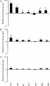
Figure 4. Quantification of p21 and CyclinD1 mRNA
mRNA for the cell-cycle markers p21 (A) and CyclinD1 (B) and the 28S rRNA (C) after 30 (T30) and 90 (T90) days of training and following 3 (D3), 10 (D10), 30 (D30), 60 (D60) and 90 (D90) days of detraining. The RNA levels are normalized to GAPDH mRNA and are relative to the pre-training value for each subject. * Significantly different from pre-training value (pre-training value = 1); # significantly different from T90.
Figure 5. Myonuclear number
The mean number of myonuclei per muscle fibre before training (Pre), after 30 (T30) and 90 (T90) days of training and following 3 (D3), 10 (D10), 30 (D30), 60 (D60) and 90 (D90) days of detraining.
Figure 6. Fibre area
Mean cross-sectional area of muscle fibres before training (Pre), after 30 (T30) and 90 (T90) days of training and following 3 (D3), 10 (D10), 30 (D30), 60 (D60) and 90 (D90) days of detraining. * Significantly different from Pre; # significantly different from T90.
Figure 7. Myonuclear domain
Myonuclear domain before training (Pre), after 30 (T30) and 90 (T90) days of training and following 3 (D3), 10 (D10), 30 (D30), 60 (D60) and 90 (D90) days of detraining. * Significantly different from Pre; # significantly different from T90.
Similar articles
- The effect of resistance training, detraining and retraining on muscle strength and power, myofibre size, satellite cells and myonuclei in older men.
Blocquiaux S, Gorski T, Van Roie E, Ramaekers M, Van Thienen R, Nielens H, Delecluse C, De Bock K, Thomis M. Blocquiaux S, et al. Exp Gerontol. 2020 May;133:110860. doi: 10.1016/j.exger.2020.110860. Epub 2020 Feb 1. Exp Gerontol. 2020. PMID: 32017951 - Enhanced satellite cell proliferation with resistance training in elderly men and women.
Mackey AL, Esmarck B, Kadi F, Koskinen SO, Kongsgaard M, Sylvestersen A, Hansen JJ, Larsen G, Kjaer M. Mackey AL, et al. Scand J Med Sci Sports. 2007 Feb;17(1):34-42. doi: 10.1111/j.1600-0838.2006.00534.x. Scand J Med Sci Sports. 2007. PMID: 17305939 - Assessment of satellite cell number and activity status in human skeletal muscle biopsies.
Mackey AL, Kjaer M, Charifi N, Henriksson J, Bojsen-Moller J, Holm L, Kadi F. Mackey AL, et al. Muscle Nerve. 2009 Sep;40(3):455-65. doi: 10.1002/mus.21369. Muscle Nerve. 2009. PMID: 19705426 - The behaviour of satellite cells in response to exercise: what have we learned from human studies?
Kadi F, Charifi N, Denis C, Lexell J, Andersen JL, Schjerling P, Olsen S, Kjaer M. Kadi F, et al. Pflugers Arch. 2005 Nov;451(2):319-27. doi: 10.1007/s00424-005-1406-6. Epub 2005 Aug 10. Pflugers Arch. 2005. PMID: 16091958 Review. - The satellite cell as a companion in skeletal muscle plasticity: currency, conveyance, clue, connector and colander.
Anderson JE. Anderson JE. J Exp Biol. 2006 Jun;209(Pt 12):2276-92. doi: 10.1242/jeb.02088. J Exp Biol. 2006. PMID: 16731804 Review.
Cited by
- Fast and slow myofiber nuclei, satellite cells, and size distribution with lifelong endurance exercise in men and women.
Montenegro CF, Skiles C, Kuszmaul DJ, Gouw A, Minchev K, Chambers TL, Raue U, Trappe TA, Trappe S. Montenegro CF, et al. Physiol Rep. 2024 Jul;12(13):e16052. doi: 10.14814/phy2.16052. Physiol Rep. 2024. PMID: 38987200 Free PMC article. - Impact of a 16-week strength training program on physical performance, body composition and cardiac remodeling in previously untrained women and men.
Grandperrin A, Ollive P, Kretel Y, Maufrais C, Nottin S. Grandperrin A, et al. Eur J Sport Sci. 2024 Apr;24(4):474-486. doi: 10.1002/ejsc.12033. Epub 2024 Mar 18. Eur J Sport Sci. 2024. PMID: 38895874 Free PMC article. - Combinatorial metabolomic and transcriptomic analysis of muscle growth in hybrid striped bass (female white bass Morone chrysops x male striped bass M. saxatilis).
Rajab SAS, Andersen LK, Kenter LW, Berlinsky DL, Borski RJ, McGinty AS, Ashwell CM, Ferket PR, Daniels HV, Reading BJ. Rajab SAS, et al. BMC Genomics. 2024 Jun 10;25(1):580. doi: 10.1186/s12864-024-10325-y. BMC Genomics. 2024. PMID: 38858615 Free PMC article. - Sex Hormones and Satellite Cell Regulation in Women.
Oxfeldt M, Dalgaard LB, Farup J, Hansen M. Oxfeldt M, et al. Transl Sports Med. 2022 Apr 14;2022:9065923. doi: 10.1155/2022/9065923. eCollection 2022. Transl Sports Med. 2022. PMID: 38655160 Free PMC article. Review. - Molecular and Structural Alterations of Skeletal Muscle Tissue Nuclei during Aging.
Cisterna B, Malatesta M. Cisterna B, et al. Int J Mol Sci. 2024 Feb 2;25(3):1833. doi: 10.3390/ijms25031833. Int J Mol Sci. 2024. PMID: 38339110 Free PMC article. Review.
References
- Adams GR. Role of insulin-like growth factor-I in the regulation of skeletal muscle adaptation to increased loading. In: Holloszy JO, editor. Exercise and Sport Sciences Reviews. Baltimore: Williams & Wilkins; 1998. - PubMed
- Adams GR, Haddad F, Baldwin KM. Time course of changes in markers of myogenesis in overloaded rat skeletal muscles. J Appl Physiol. 1999;87:1705–1712. - PubMed
- Allen DL, Monke SR, Talmadge RJ, Roy RR, Edgerton VR. Plasticity of myonuclear number in hypertrophied and atrophied mammalian skeletal muscle fibers. J Appl Physiol. 1995;78:1969–1976. - PubMed
- Allen DL, Roy RR, Edgerton VR. Myonuclear domains in muscle adaptation and disease. Muscle Nerve. 1999;22:1350–1360. - PubMed
Publication types
MeSH terms
LinkOut - more resources
Full Text Sources
Research Materials


